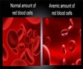
University of Copenhagen researchers in Denmark have developed a new X-ray imaging technique that can capture the three-microsecond process whereby haemoglobin molecules in red blood
A new X-ray imaging technique that can capture the three-microsecond process whereby haemoglobin molecules in red blood cells give up oxygen to keep the body alive has been developed by University of Copenhagen researchers in Denmark.
This process //involves a haemoglobin molecule's transformation from a "relaxed" form, which can bond to oxygen, to a "tense" form, which squeezes out the oxygen.The researchers say that their imaging technique offers a way to image proteins in their natural, fast-moving state.
Scientists traditionally used X-ray crystallography to image protein structures - a process that required making the structures into rigid crystals, firing X-ray beams at them, and recording the geometric patterns produced.
Though the procedure helps work out the structure of DNA, it provides little evidence of what structures materials have in their natural setting.
Research leader Marco Cammarata and his colleagues wanted to transfer X-ray techniques to free-moving proteins in solution, but individual molecules in this setting tend to reflect too few X-rays to provide a clear picture.
The researchers say that the solution could be to use brute force - a X-rays from the most powerful source available, a kind of particle accelerator called a synchrotron.
Advertisement
The researchers conducted their study at the European Synchrotron Radiation Facility in Grenoble, France.
Advertisement
Cammarata says that the X-rays that "illuminate" the protein can be analysed to work out its 3D structure.
He adds that firing short pulses captures the protein's motion, just like a fast exposure on a normal camera will freeze a moving person.
The researchers claim that they have already found evidence of a previously unknown haemoglobin structure, which it adopts for only a short time as the protein gives up its oxygen.
They now plan to study other, less-common proteins.
"Applying this technique to little known protein systems and to the study of (intermediate forms) is where we will make a really important contribution," New Scientist magazine quoted Cammarata as saying.
A research article on the new process has been published in the journal Nature Methods.
Source-ANI
RAS/SK


