‘Bone in a dish’ is an artificially engineered bone construct that contains bone cells derived from stem cells embedded within the structure. It allows the studying of cancer and bone healing more closely.
- A new technology known as ‘bone in a dish’ has been developed
- It consists of an artificially engineered mineralized structure with embedded stem cells
- This novel technology facilitates research in the area of cancer and bone regeneration
This new ‘bone in a dish’ technology is being used to closely study various disease processes, including the initiation and progression of bone cancer, as well as for developing therapies for bone injury.
“What is remarkable is that researchers in our field have become used to cultivating cells within a protein mixture to approximate how cells live in the body. But this is the first time anyone has been able to embed cells in minerals, which is what characterizes the bone tissue,” said Dr. Luiz E. Bertassoni, who led the study.
The study has been published in Nature Communications, a constituent journal of the Nature Publishing Group.
Read More..
Study Team
The study was led by Dr. Luiz E. Bertassoni, DDS, PhD, who is an Associate Professor at the Oregon Health & Science University (OHSU) in Portland, Oregon, USA. He holds joint appointments at the Department of Restorative Dentistry, the OHSU Center for Regenerative Medicine, Department of Biomedical Engineering and the Cancer Early Detection Advanced Research Center (CEDAR) at the Knight Cancer Institute.Other team members included Dr. Greeshma Thrivikraman Nair, PhD, who is a postdoctoral fellow at the OHSU School of Dentistry and Avathamsa Athirasala, BTech, MS, who is a PhD student in Bertassoni’s lab.
How was the ‘Bone in a Dish’ Constructed?
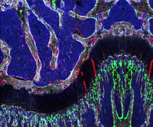
The new technology, which resembles a miniaturized ‘bone in a dish,’ requires just 72 hours for preparing and becoming functional. It was constructed by mixing human stem cells with a solution of collagen – a common protein available abundantly, which is an essential component of bone tissue matrix. The collagen proteins become linked together, forming a gel, consisting of a mesh-like network in which the stem cells become embedded. This gel containing embedded stem cells was then exposed to a solution containing dissolved calcium and phosphate, which are essential minerals present in bone.Another essential component of this mixture was osteopontin, which is a phosphorylated sialic acid-rich non-collagenous protein present in bone matrix. Osteopontin slows down the process of crystal formation by binding to the calcium and phosphate molecules. This binding also reduces cellular toxicity caused by these minerals. The whole mixture percolates through the spongy collagen matrix and the minerals gradually form ordered layers of crystals that resemble actual bone.
“We can reproduce the architecture of bone down to a nanometer scale,” says Bertassoni. “Our model goes through the same biophysical process of formation that bone does.”
How was the ‘Bone in a Dish’ Technology Used to Study Disease Models?
The stem cells embedded within the mineralized collagen matrix developed into fully functional bone cells, osteoblasts and osteocytes, without the need for any additional nutrients or growth factors. Subsequently, these cells developed the capacity to connect and communicate with neighboring cells. Thus, the artificially engineered bone-like structure provided a microenvironment similar to that present in actual bone, which was ideal for the growth and differentiation of the stem cells into bone cells. This microenvironment also allowed nerve cells and endothelial cells to thrive and develop interconnected networks within the mineralized structure.The engineered bone was evaluated in vivo in a mouse model of prostate cancer. The mineralized structure was implanted beneath the skin of mice and in due course, blood vessels developed from the embedded stem cells, which interconnected with the natural blood vasculature of the mice. Upon injection of prostate cancer cells within the vicinity of the mineralized bone implant, it was observed that the tumor growth was three times higher than in control mice that didn’t have the mineralized bone implant.
What are the Potential Applications of the ‘Bone in a Dish’ Technology?
The major application of the new technology will be in the area of disease modeling. For example, this technology will be ideal for closely studying bone function, bone regeneration, as well as various aspects of cancer such as initiation, growth, and metastasis. In fact, this novel technology could potentially transform the field of Regenerative Medicine by replacing the need for autologous bone transplants, which are currently used to regenerate injured bone.Future Plans
The research team is planning to engineer a new variant of the mineralized artificial bone with embedded bone marrow cells to study various aspects of leukemia, such as initiation, development and metastasis. Moreover, they are in the process of testing the artificially engineered bone as a replacement for damaged bone in animal models.Funding Source
The research was funded by the National Institute of Dental and Craniofacial Research of the National Institutes of Health (NIH), the American Academy of Implant Dentistry Foundation, the OHSU-PSU Collaboration Project Seed Funding, the OHSU Knight Cancer Institute, the Pacific Northwest National Laboratory (PNNL) under the US Department of Energy (DOE), and the National Science Foundation, USA.Source-Medindia


