Dengue infections can now be monitored in real time using positron emission tomography to identify effectiveness of treatment.
- Positron Emission Tomography (PET) Imaging is used extensively for cancer diagnosis but is now being considered an ‘old’ and outdated method of diagnosis
- A research team from Duke University, Singapore, has utilized PET for dengue infection imaging
- This is the first time that this imaging technique has been used to study status and progression of dengue infection
- As an imaging tool to identify and trace the progress of dengue infections in real time
- To determine the effectiveness of a dengue treatment program
Mechanism of Function
In general PET, a radioactive material is either injected or inhaled into the body, which then accumulates in the organ or tissues. The emissions from the radiotracer are detected by a special camera and monitored on a computer. FDG is a radioactive version of glucose that can be used to track where the cells take up body glucose.In the current study, the FDG was injected into mice. The research team knew that dengue infected mice had inflammation of the large and small intestines; this was used as a marker for detection of dengue. Inflammation resulted in an increased uptake of glucose by the cells in the intestines, which could be visualized by the PET-FDG technique.
Dr. Ann-Marie Chacko who is the lead author of the study and an Assistant Professor of Duke-NUS said that this was the first time that PET was used to determine the presence of an acute viral infection. This finding can be used to obtain quality images of dengue infection in the body using non-invasive technology.
The findings of the study included:
- Inflammation was detected in the small, large intestines and the spleen of dengue infected mice
- After anti-viral therapy was administered to the mice, the inflammation subsided
- The severity of the infection could be predicted by assessing glucose uptake by the FDG-PET
The viral load present in the blood has been the important method of measuring the severity of infection, according to Dr. Jenny Low, a senior consultant at Duke-NUS. The scientist revealed that the major factor in a clinical trial is the absence of an effective measure of treatment, which this imaging technique could solve. The various potential treatment procedures used in clinical trials for dengue can now be monitored in real time using this FDG-PET procedure, identifying the effectiveness of the treatment.
Dengue Infections
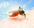
Dengue esults in flu-like symptoms and is a mosquito borne viral infection. It is an infectious disease which occurs in tropical and sub-tropical regions in the world. There has been an increase in the global incidence of dengue,and, since there is no cure for dengue, treatment is focused on lowering the intensity of symptoms.Early detection of dengue is very important as it helps in lowering fatality to 1%. A lot of research is being conducted on dengue infections to identify a potential cure for the disease, although, there is no known cure till now. The PET imaging technique could be used to determine the presence of the dengue infection and for monitoring the progression of the disease, irrespective of the severity of the symptoms. This will help to identify high risk patients or can be used to determine if the infection has truly subsided or not.
References:
- Positron Emission Tomography - Computed Tomography (PET/CT) - (https://www.radiologyinfo.org/en/info.cfm?pg=pet)
- Dengue and severe dengue - (http://www.who.int/mediacentre/factsheets/fs117/en/)
- Positron Emission Tomography - Computed Tomography (PET/CT) - (https://www.ncbi.nlm.nih.gov/pmc/articles/PMC5414563/)


