Magnetic resonance imaging (MRI) can help monitor liver fat in obese patients. This is done by a technique called chemical shift encoded MRI (CSE-MRI) for measuring the proton density fat fraction (PDFF).
- A variation of the magnetic resonance imaging (MRI) technique called chemical shift encoded MRI (CSE-MRI) can monitor liver fat in obese patients
- This is done by measuring a liver fat fraction called proton density fat fraction (PDFF)
- This tool will be of great benefit to obese patients with fatty livers suffering from conditions such as non-alcoholic fatty liver disease (NAFLD)
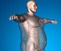
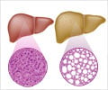
Obesity is a major public health problem in Western countries like the US. Approximately two-thirds of adults in the USA are either overweight or obese. Obesity is associated with fat accumulation in the liver in conditions such as non-alcoholic fatty liver disease (NAFLD). The disease can progress to non-alcoholic steatohepatitis (NASH) and liver cirrhosis, which significantly increases the risk of liver cancer.
Weight reducing surgical procedures such as bariatric surgery and sleeve gastrectomy are very effective in reducing body fat. However, whether overall body fat reduction also causes a decrease in liver fat is yet to be established. The only available procedure is a biopsy, followed by fat detection. However, liver biopsy is a highly invasive procedure and is not at all suitable for routine diagnosis and monitoring of progression of fatty liver diseases.
Read More..
What’s New in the Current Study?
The new study used a noninvasive technique called chemical shift encoded magnetic resonance imaging (CSE-MRI) to quantitatively estimate the liver fat content before and after weight loss bariatric surgery. The procedure assesses the fat content of the liver by measuring a fat fraction called proton density fat fraction (PDFF).Pooler indicated that CSE-MRI allows the representation of the liver fat content as a percentage, which is much easier for the patients to comprehend. The percentage value also permits the direct comparison of the fat content of liver from surgical as well as biopsy samples, without any confusion or ambiguity.
What is CSE-MRI?
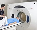
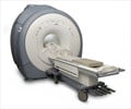
Chemical shift encoded magnetic resonance imaging (CSE-MRI) is an adaptation of the MRI technique, which is a noninvasive imaging method using a powerful magnetic field, radio waves and a computer to produce detailed images of the internal structures of the body. The images produced by MRI are clearer and much more detailed than other imaging techniques.CSE-MRI takes advantage of the difference between the properties of water and fat in an organ. The differences in the local magnetic field produced by variation in the fat density due to presence of water molecules, helps to quantitatively assess fat composition of the body (in the present study, fat content of liver) and estimate the degree of saturation of the fat (saturated, monounsaturated, or polyunsaturated), thereby producing very accurate, clear-cut images of the liver.
When PDFF is measured by CSE-MRI, it helps to detect the presence of fatty liver. Thus, PDFF can act as an accurate biomarker of fatty liver diseases such as non-alcoholic fatty liver disease (NAFLD).
Study Methods
The study involved CSE-MRI to assess the condition of 50 obese patients both before and after undergoing bariatric surgery for weight reduction. The patients were put on a very low-calorie diet (VLCD) for 14 days before the surgery, which has been found to enhance the safety and efficacy of this type of surgery.Before the surgery, CSE-MRI was carried out twice, first before initiation of the VLCD and second, one-day pre-op. After the surgery, CSE-MRI was carried out thrice – at 1 month post-op, 3 months post-op, and 6 months post-op. The research team also determined liver fat by PDFF and compared it with the body mass index (BMI) and waist circumference, which are important physical markers of obesity.
Study Findings
The following findings concerning the study participants should be noted:- Six to 10 months after the surgery, the mean PDFF dropped from 18 percent to 5 percent (normal range is 5% or less)
- During the same time, the mean BMI decreased from 45 to 34.5
- The PDFF normalized at approximately five months following the surgery
"The results showed a rapid early phase of improvements in liver fat, followed by a phase of continued improvements at a slower pace," Pooler said. "The changes began with the initiation of the low-calorie diet and occurred in advance of the overall improvements in BMI among the patients."
Study Implications
The following implications were noted, with reference to the role of CSE-MRI in the management of obese patients suffering from fatty livers:- Measurement of PDFF could help to select patients for bariatric surgery since there is a strong correlation between reduction of liver fat and pre-op liver fat content.
- It would be helpful to monitor liver fat by CSE-MRI post-op, independent of monitoring weight loss since liver fat reduction was weakly correlated with initial weight and total weight loss.
- Patients with fatty livers are likely to be most benefitted by CSE-MRI and PDFF estimation, regardless of their initial weight or weight loss.
Future Prospects
The research team believes that the CSE-MRI technique could have many applications, other than just monitoring the effects of bariatric surgery. The procedure could potentially monitor all types of patients who have undergone a variety of weight reduction treatments. The researchers aim to extend their study to these groups of patients in the future.Reference:
- Monitoring Fatty Liver Disease with MRI Following Bariatric Surgery: A Prospective, Dual-Center Study - (https://pubs.rsna.org/doi/10.1148/radiol.2018181134)
Source-Medindia


