Perivascular fat imaging detects coronary inflammation, an early sign of coronary artery disease, for suitable intervention to prevent future complications.
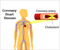
- Coronary artery inflammation represents early phase of coronary artery disease (CAD) that is potentially reversible if suitable measures are put in place on time
- Existing diagnostic methods do not accurately identify coronary inflammation
- Current study describes perivascular fat imaging as a tool to detect coronary inflammation for initiation of preventive measures and to prevent disease progression
Perivascular Fat CT Imaging Vs Current Methods To Predict CAD Risk
The authors of the study believe that perivascular fat imaging is superior to currently available methods for the following reasons- Blood markers such as C-reactive protein and proinflammatory cytokines are not directly involved in the formation of atherosclerotic plaque, or local processes and cannot detect unstable plaques
- Existing noninvasive [computerized tomography angiography (CTA) or 18F-fluorodeoxyglucose (18FFDG)–positron emission tomography (PET)] and invasive (intravascular ultrasound and optical coherence tomography) imaging tools do not provide accurate information on coronary inflammation.
- The only established noninvasive imaging biomarker of predictive value in primary prevention is Coronary calcium scoring (CCS), but it identifies only irreversible changes in the vessel wall that are not altered by interventions to reduce CAD risk, such as statins or lifestyle changes.
Perivascular Fat Measurement With Histological And PET Validation
- For the study, the team performed perivascular fat CT scans that measured FAI in 453 patients undergoing cardiac surgery, which measured adipocyte content and size.
- On perivascular imaging, higher FAI was found to be associated with greater degree of coronary inflammation (confirmed by FDG uptake on PET).
- Absence of inflammation promotes lipid accumulation within the adipocyte and allows differentiation to more larger and more mature form. This is a good sign and associated with low FAI
- Inflammation reduces lipid accumulation, and slows down differentiation to more mature adipocyte. This is associated with a higher FAI, predisposing to unstable plaques, plaque rupture and cardiac events.
- The above CT findings were validated with samples of arterial tissue obtained during surgery which were sent for histological analysis to confirm the size and fat content of the adipocytes as described above.
- Presence of coronary inflammation was also confirmed by tissue uptake of 18F-fluorodeoxyglucose in positron emission tomography, which is the gold standard method to noninvasively assess tissue inflammation in vivo.
- The team went on to measure FAI in 273 additional patients (156 with and 117 without significant coronary plaques) in a clinical setting and who underwent coronary angiography.
The findings of the study suggest that inflammation reduces perivascular fat accumulation leading to a higher FAI and local vascular stimuli play a part in these processes.
"Our heart arteries are surrounded by fat. The fat detects inflammation, becoming more watery and less fatty as it sits next to an inflamed artery," said Charalambos Antoniades, a senior author on the paper at the University of Oxford.
Inflammation And Atherogenesis (CAD) – A Crucial Link
- Inflammation is a key component in atherogenesis, and any tool that can accurately detect vascular inflammation would be ideal to detect high risk lesions and put in place appropriate interventional measures.
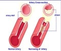
- Inflammation also characterizes unstable atherosclerotic plaques, which may rupture, leading to major cardiovascular events such as heart attacks and strokes.
- Noninvasive and early detection of vascular inflammation will be a game changer in the field of preventive cardiology, which is what this study hopes to have achieved.
Scope of The Study And Perivascular Fat Imaging Test
- The CT scan is a simple non-invasive test that can be done in an outpatient setting and picks up early subclinical CAD.
- Clinicians could detect changes in FAI to determine whether patients are responding to treatment, or make appropriate interventions in people at risk for arterial plaque rupture.
"The key advantage of this method is to be able to detect early changes in blood vessels that precede cardiovascular disease," said Caitlin Czajka, an associate editor at Science Translational Medicine.
What Does the Future Look Like?
- Clinical follow up studies to establish the predictive accuracy of FAI for cardiac events are ongoing and the authors expect to publish the findings by the end of 2017.
- Currently, although the CT test is in itself simple, the calculation of the FAI from imaging studies is a long drawn out procedure, needing a skilled technician. The authors are working to make their software more user friendly and accessible.
References:
- Noninvasive Imaging Gets to the Heart of Cardiac Disease Risk - (https://www.aaas.org/news/noninvasive-imaging-gets-heart-cardiac-disease-risk)


