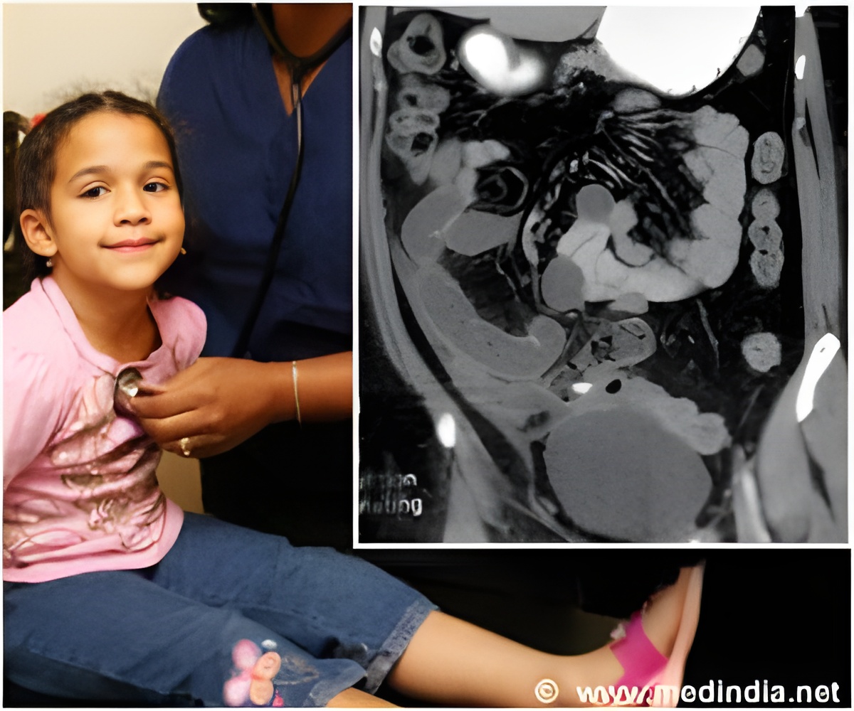
A new study found MR Enterography to be well-tolerated and practical to use amongst children with IBD.

Magnetic Resonance Enterography is commonly used to diagnose and monitor Inflammatory Bowel Disease (IBD). It is the preferred choice over Computed Tomography for evaluation of IBD as it is a non-invasive, radiation free interrogation. Although colonoscopy is usually used in the final diagnosis and detecting changes in IBD, the cost involved is high.
This is one of the first studies to evaluate the role of MRE in studying the changes in gut inflammation amongst children with spondyloarthritis.
In this small pilot study, five children, four with spondyloarthritis and one with juvenile idiopathic arthritis were identified for sub-clinical inflammatory bowel disease diagnosed using the fecal test.
Three children were identified with signs of IBD. The findings included –
• thickening and uptake of contrast agent at the distal end of the small intestine in one child;
• prominent vasa recta (intestinal arteries) and mesenteric lymph nodes (lymph nodes that lie between the lining of the abdominal cavity connecting the duodenum and small intestine to the back wall of the abdomen) in the third child.
The researchers found that the use of MRE amongst children with sub-clinical inflammatory bowel disease was well-tolerated and practical. It shows the possibility of the use of this test to screen sub-clinical bowel disease.
Reference: MR enterography to evaluate sub-clinical intestinal inflammation in children with spondyloarthritis; Mathew Stoll et al; BMC Pediatric Rheumatology 2012.
Source-Medindia
 MEDINDIA


 Email
Email


