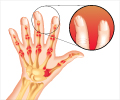
Novel positron emission tomography (PET) imaging method that more fully evaluates the extent of rheumatoid arthritis inflammation has been developed by a research team. The new method is designed to target translocator protein (TSPO) expression in the synovium.
- A new imaging technique shows the nature of inflammation in rheumatoid arthritis.
- The new technique is a positron emission tomography (PET) that targets translocator protein (TSPO) expression in the synovium.
- The synovium is a connective tissue in the joints.
She points out, "Here we present the first ever analysis of TSPO expression in the major constituents of RA pannus (inflamed synovium), demonstrating that TSPO PET likely acts as an imaging tool of not only macrophages, but also activated synovial fibroblasts, a cell group increasingly recognized to play a critical role in RA inflammation."
The study included three RA patients and three healthy volunteers who underwent PET scans of both knees using the TSPO radioligand carbon-11 (11C)-PBR28. In addition, cellular expression of TSPO was examined in synovial tissue from these individuals, plus three more RA patients and three more healthy patients (undergoing knee arthroscopy for injuries). TSPO mRNA expression and hydrogen-3 (3H)-PBR28 radioligand binding were assessed using in vitro monocytes, macrophages, fibroblast-like synoviocytes (FLS) and CD4+ T-lymphocytes.
Results showed that the 11C-PBR28 PET signal was significantly higher in RA joints compared to healthy joints. In addition, 3H-PBR28 specific binding in synovial tissue was approximately 10-fold higher in RA patients compared to healthy controls. Immunofluorescence revealed TSPO expression on macrophages, FLS and CD4+ T cells. In vitro study demonstrated highest TSPO mRNA expression and 3H-PBR28 specific binding in activated FLS, non-activated and activated 'M2' reparative macrophages. The lowest TSPO expression was in activated and non-activated CD4+ T lymphocytes.
Narayan notes, "It is well recognized that not all currently available treatments are capable of controlling joint inflammation in all patients with rheumatoid arthritis, hence the need to develop new pharmacological therapies. This work demonstrates that TSPO PET is able to act as a means of imaging not only synovial macrophages, but also activated synovial fibroblasts. The crucial role of the fibroblast and its soluble products in RA pathogenesis is increasingly realized."
References:
- Translocator Protein as an Imaging Marker of Macrophage and Stromal Activation in Rheumatoid Arthritis Pannus - (http://jnm.snmjournals.org/content/59/7/1125)


