Study shows that red color channels help in better detection of cysts in cystoid macular edema especially in the ethnic minority.

- In racial/ethnic minorities, who normally have darker fundus, cysts, that result from cystoid macular edema usually goes undetected.
- New study states that inspecting the red color channels from the color photographs of the fundus that are used in screening diabetes-related eye disease, can improve the ability to detect diabetic macular edema, especially in the racial/ethnic minorities community.
- This is because the red color channels have long wavelength and help in detailed view of the vessels in the intermediate and deeper layers of the retina.
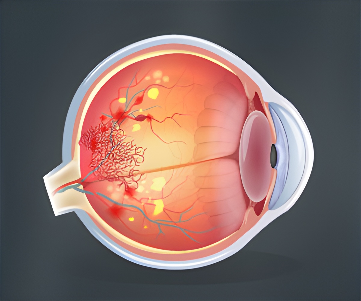
Diabetic macular edema is a complication of diabetes in which there is fluid accumulation in the retina-structure in the back of the eye that reflects light.
This results from leaky blood vessels in the retina. This condition can lead to blindness among working-age adults with diabetic eye disease. It affects approximately 21 million people worldwide.
In this study, researchers focused on cystoid macular edema.
It is a major cause of severe central vision loss in which a degenerative change of the neural retina occurs.
For screening diabetic eye disease color fundus photography has long been the standard method.
The lead author of the new study was Mastour A. Alhamami, PhD, of Indiana University School of Optometry, Bloomington.
Comparison of Red and Green Color Channels
For the study, researchers analyzed standard color fundus photographs obtained from 2,047 adult patients with diabetes, but who were medically under-served mostly without access to routine eye care.
Diabetics should undergo dilated eye examinations annually or at least once in two years, to detect early signs of diabetic eye disease.
But most participants admitted to not have undergone in at least three years.
Among the patients, 90% belonged to racial/ethnic minorities (other than non-Hispanic white).
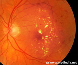
The retinal photographs showed clinically significant macular edema in 148 patients.
Of these, 13 patients had a cystoid pattern of macular edema.
The photographs were divided into red color channels with long wavelength and green color channels with short wavelength and then compared.
The results showed that 12 of the 13 patients with cystoid macular edema had a dark-colored fundus.
Macular cysts were easier to detect when viewed using the red-channel images, compared to the green-channel images as green-channels have shorter wavelength and does not allow clear examination of the deeper structures of the retina.
"Cysts may be under-detected with the present fundus camera methods, particularly when short-wavelength light is emphasized or in patients with dark fundi," Dr. Alhamami said.
This new findings offer a clear-cut advantage of red-channel images over the green-channel images especially in under-served groups in a high proportion of people are dark-eyed and they have higher rates of diabetic eye disease.
Researchers also state that red-channel images may also provide more information about damage in the deeper layers of the retina, due to its longer wavelength.
The study is published in the journal Optometry and Vision Science.
Reference
- Mastour A. Alhamami et al. Comparison of Cysts in Red and Green Images for Diabetic Macular Edema. Optometry and Vision Science ; (2017) doi: 10.1097/OPX.0000000000001010
Source-Medindia


