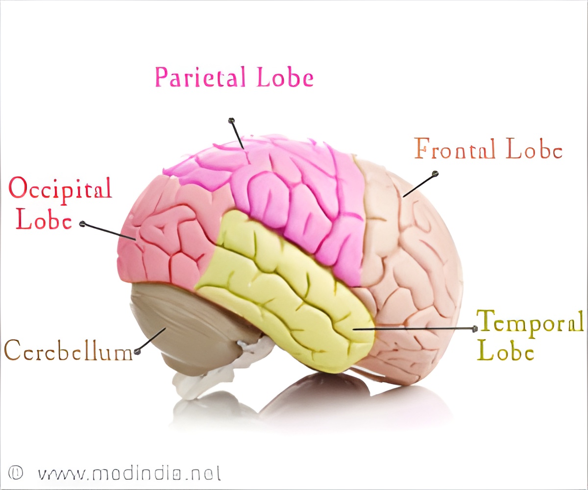
Novel, non-invasive imaging-based methods have been discovered to investigate an important structure of the human brain.

‘Novel, non-invasive imaging-based methods have been discovered to investigate the visual sensory thalamus, an important structure of the human brain and point of origin of visual difficulties in diseases such as dyslexia and glaucoma.’


The visual sensory thalamus is known to be a key region that connects the eyes with the cerebral cortex. It is the point of origin of visual difficulties in many diseases such as dyslexia and glaucoma.Decoding Hidden Territory
The area contains two tiny major compartments that are located very deep inside the brain. This made it difficult to be assessed so far, thereby adding to the struggle to decode visual sensory processing.
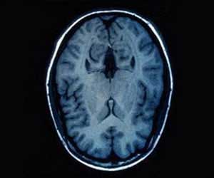
However, the study team used unique neuroimaging data with high spatial resolution on a specialized magnetic resonance imaging (MRI) machine at the MPI-CBS in Leipzig to explore the area.
It was found that the two major compartments of the visual sensory thalamus are characterized by different amounts of brain white matter (myelin), as detected in novel MRI data, obtained from post-mortem studies of developmental dyslexia.
Advertisement
Source-Medindia


