Physicians are seeking more powerful drugs to subdue strains of pathogenic bacteria that are resistant to the antibiotics that fought against their predecessor generations.
The team will present their findings at the annual meeting of the American Crystallographic Association, held July 28 – Aug. 1 in Boston, Mass.
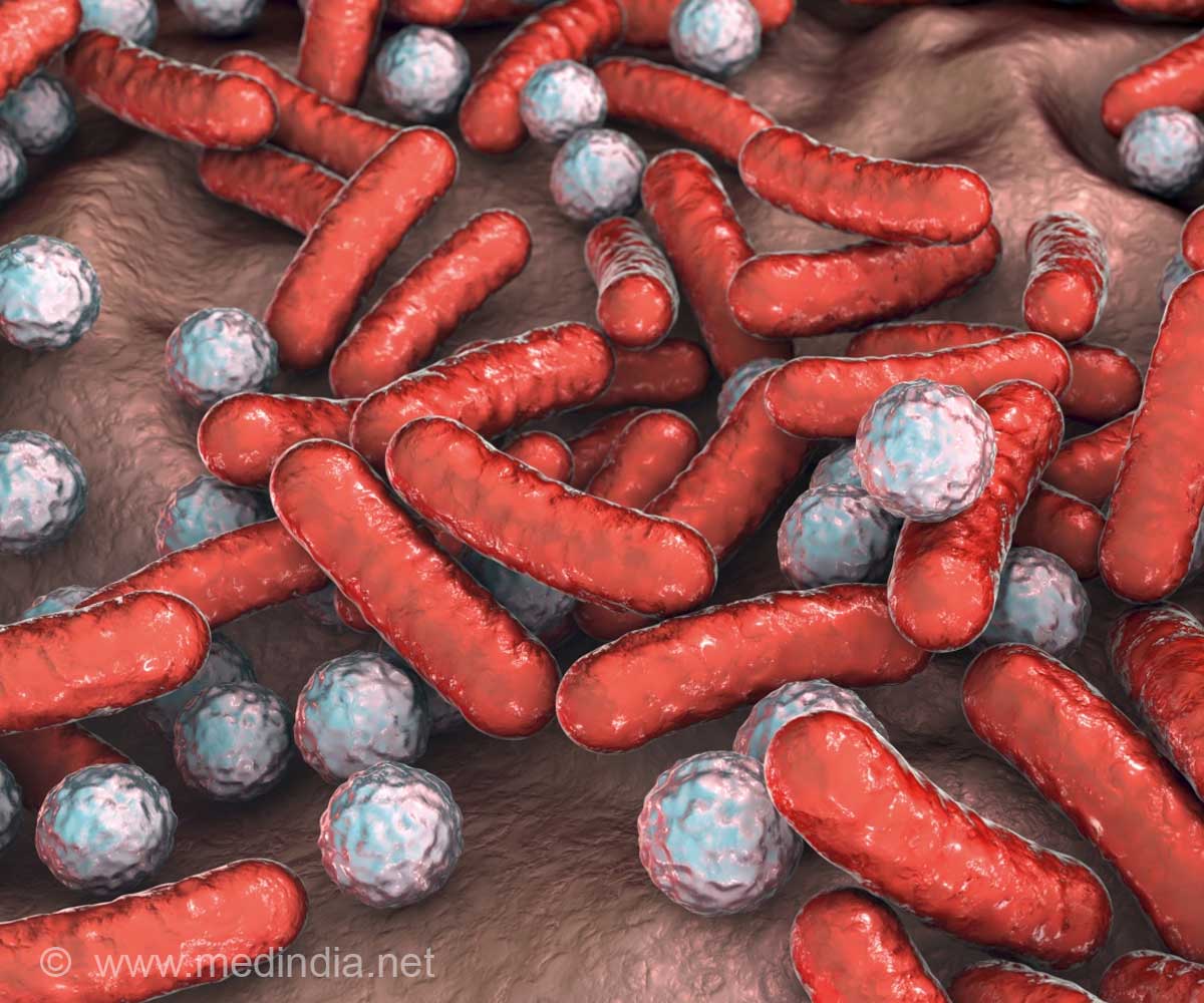
For years, the drug vancomycin has been the last-stand treatment for life-threatening cases of methicilin-resistant Staphylococcus aureus, or MRSA. A powerful antibiotic first isolated in 1953 from soil collected in the jungles of Borneo, vancomycin works by inhibiting formation of the S. aureus cell wall so that it cannot provide structural support and protection. In 2002, however, a strain of S. aureus was isolated from a diabetic kidney dialysis patient that would not succumb to vancomycin. This was the first recorded instance in the United States of vancomycin-resistant Staphylococcus aureus, or VRSA, a deadly variant that many now consider one of the most dangerous bacteria in the world.
Former UNC graduate student Jonathan Edwards (now at the Massachusetts Institute of Technology), under the guidance of chemistry professor Matthew Redinbo, led the research team that sought a detailed biochemical understanding of the VRSA threat. They focused on a S. aureus plasmid—a circular loop of double-stranded DNA within the Staph cell separate from the genome—called plW1043 that codes for drug resistance and can be transferred via conjugation ("mating" that involves genetic material passing through a tube from a donor bacterium to a recipient).
Before the plasmid gene for drug resistance can be passed, it must be processed for the transfer. This occurs when a protein called the Nicking Enzyme of Staphylococci, or NES, binds with its active area, known as the relaxase region, to the donor cell plasmid. NES then cuts, or "nicks," one strand of the double helix so that it separates into two single strands of DNA. One moves into the recipient cell while the other remains with the donor. After the two strands are replicated, NES reforms the plasmid in both cells, creating two drug-resistant Staph that are ready to spread their misery further.
Using x-ray crystallography, Edwards, Redinbo and their colleagues defined the structure of both ends of the VRSA NES protein, the N-terminus where the relaxase region resides and the molecule's opposite end known as the C-terminus. They noticed that the N-terminus structure included a region with two distinct protein loops. Suspecting that this area might play a critical role in the VRSA plasmid transfer process, the researchers cut out the loops. This kept the NES relaxase region from clamping onto or staying bound to the plasmid DNA.
Advertisement
"We realized that a compound that could block this groove, prevent the NES loops from attaching and inhibit the cleaving of the plasmid DNA into single strands could potentially stop conjugal transfer of drug resistance altogether," Edwards says.
Advertisement
Redinbo says that this "proof of concept" experiment suggests that the same inhibition might be possible in vivo. "Perhaps by targeting the DNA minor groove, we might make antibiotics more effective against VRSA and other drug-resistant bacteria," he says.
Source-Eurekalert


