A new research technique changes open the possibility of helping identify new ways to protect dental tissues from acids and develop new treatments.

This enabled them to build clear 3D images of the tooth’s internal structure with sub-micrometer resolution (a micrometer being one-thousandth of a millimeter).
By analyzing these images over the six hours of the experiment, researchers conducted the first-ever time-resolved 3D study (often referred to as 4D studies) of the microstructural changes caused by acid in teeth and published in Dental Materials.
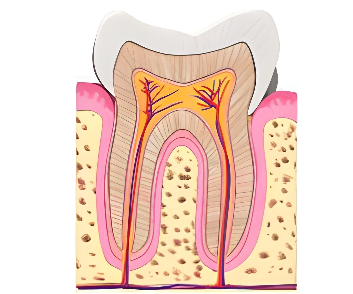
Dentine is a part of human teeth that makes tooth surface strong and resilient, but acids from dental plaque can cause tooth decay which affects the integrity of the dental structure.
This new technique aims to develop knowledge that leads to new treatments that can restore the structure and function of dentine.
Researchers will continue to study the mechanical response of dentine to masticatory forces in correlation with the microstructural changes that acid causes as well as in response to different treatments like fillings and crowns.
Advertisement
Source-Medindia


