Researchers discovered a new gene therapy that turns glial brain cells into neurons for restoring visual function in stroke-affected mice models. This offers hope to restore motor function affected in stroke.

‘Restoration of visual function after stroke through neurod1-mediated gene therapy was demonstrated.’


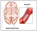
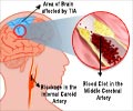
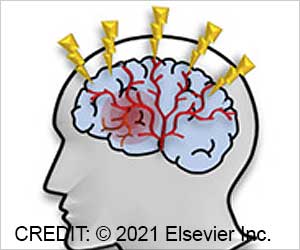
Stroke affects motor function as major arteries are mostly located in the brain. It can also affect vision, causing patients to lose their vision or find it compromised or diminished. Nerve cells don’t regenerate but the brain can sometimes remap its neural pathways to restore some visual function after a stroke. This process is slow, inefficient, and for some patients, it never happens at all.
In such cases, stem cell therapy can help. Stem cell therapy relies on finding an immune match and is cumbersome and difficult. Whereas new gene therapy as demonstrated in a mouse model is more efficient and much more promising.
There is no need to implant new cells, so there’s no immunogenic rejection. Gene therapy is easier to do than stem cell therapy and causes less damage to the brain.
Gene therapy helps the brain heal itself. The connections between the old neurons and the newly reprogrammed neurons can get re-established to restore the vision .
Advertisement
Using adeno-associated viruses they delivered the NeuroD1 gene to glial cells in the affected part of the brain. Later the glial cells were reprogrammed into neurons and were integrated into the visual cortex.
Advertisement
Understanding this technique can lead to a similar technique re-establishing motor function. This study also bridges the gap in understanding between the basic interpretation of the neurons and the function of the organs.
Source-Medindia

