Specific immune cells known as macrophages have the ability to secrete or produce a protective or healing factor known as Interleukin-10 (IL-10).
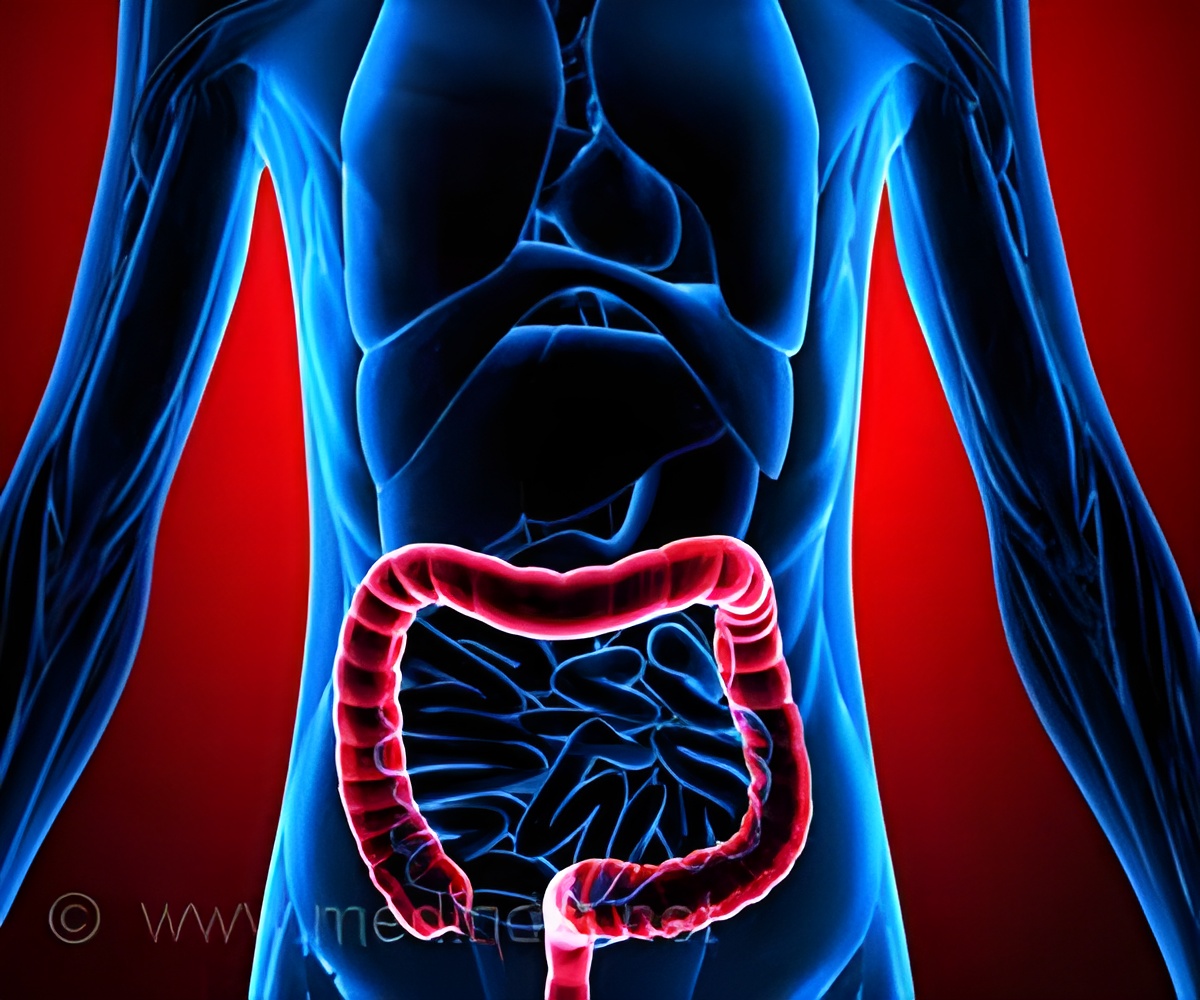
‘Specific immune cells have the ability to produce a healing factor that can promote wound repair in the intestine, and it could be a potential therapeutic avenue for the treatment of inflammatory bowel disease.’


In this study, the researchers found that a specific population of immune cells called macrophages have the ability to secrete or produce a protective or healing factor known as Interleukin-10 (IL-10), which can interact with receptors on intestinal epithelial cells to promote wound healing. The findings are published in The Journal of Clinical Investigation. "Understanding how wounds can be healed is believed to be very important and a potential therapeutic avenue for the treatment of inflammatory bowel disease," said Dr. Tim Denning, associate professor in the Institute for Biomedical Sciences at Georgia State. "In this study, we tried to understand some of the cellular mechanisms that are required for optimal wound healing in the intestine. To do this, we used a cutting-edge system, a colonoscope with biopsy forceps, to create a wound in mice. This is analogous to colonoscopies in humans. This cutting-edge system allowed us to begin to define what cells and factors contribute to wound healing in the mouse model."
The researchers used a small, fiber optic camera and forceps to pinch the mouse's intestine and take a small biopsy, just as how colonoscopies are done in humans. This small pinch created a wound, which the researchers observed as it healed. The study compared intestinal wound healing in two groups of mice: 1) typical mice (wild type) found in nature and 2) mice genetically deficient in the healing factor IL-10, specifically in macrophages, which impairs their ability to have normal wound repair.
The team also analyzed the effects of IL-10 on epithelial wound closure in vitro using an intestinal epithelial cell line.
They concluded that macrophages are a main source of IL-10 in the wound bed, and IL-10 stimulates in vitro intestinal epithelial wound healing and increases in expression during in vivo intestinal epithelial wound repair. In vitro, exposure to IL-10 increased wound repair within 12 hours and the response was further enhanced after 24 hours.
Advertisement
In addition, the researchers defined some of the signaling pathways that IL-10 uses to orchestrate wound repair. They found IL-10 promotes intestinal epithelial wound repair through the activation of cAMP response element-binding protein (CREB) signaling at the sites of injury, followed by synthesis and secretion of the WNT1-inducible signaling protein 1 (WISP-1).
Advertisement
Source-Eurekalert


