Immune cells of the brain called the “microglia” consume a voracious amount of glucose (sugar molecule) during the advent of neurodegenerative diseases.

‘Immune cells of the brain called the “microglia” consume a voracious amount of glucose (sugar molecule) during the advent of neurodegenerative diseases.
’


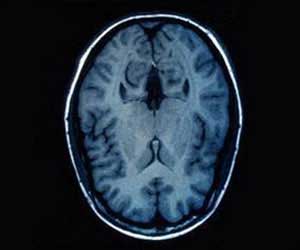
“FDG-PET” – a special variant of positron emission tomography (PET – an imaging study) is used to explore the distribution of glucose in the brain by administering an aqueous radioactive glucose solution that distributes in the brain. “Energy metabolism can be recorded indirectly via the distribution of glucose in the brain. Glucose is an energy carrier. It is therefore assumed that where glucose accumulates in the brain, energy demand and consequently brain activity is particularly high,” says Dr. Matthias Brendel, deputy director of the Department of Nuclear Medicine at LMU Klinikum München.
Microglia in Neurodegeneration
It is generally assumed that FDG-PET signal during brain imaging stems predominantly from brain cells –neurons, as they are reported as the largest energy consumers of the brain.
However, the study presents a contrasting view to the existing notion and demonstrates the predominance of the microglia in consuming high sugars at the early stages of neurodegenerative disease.
Advertisement
These imaging findings may thus help in serving as a biomarker to capture the microglial response to therapeutic interventions of dementia.
Advertisement
Source-Medindia

