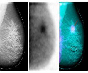
A new study shows that tomosynthesis (3D mammography) is better able to show infiltrating ductal carcinoma than 2D mammography in women at increased risk of breast cancer.

"We found that 41% (23/56 cancers) were better seen on tomosynthesis and 4% (2/56) were only seen on tomosynthesis," said Dr. Sarah O'Connell, a lead author of the study. Thirty percent of cancers (17/56) were better seen on 2D mammography but none were only seen on 2D mammography. The remaining were rated by the breast imaging specialists as being equally visible on both 2D and 3D imaging, she said.
"The majority of cancers seen better or only on tomosynthesis were predominantly infiltrating ductal carcinoma, which typically presents as a mass, focal asymmetry or architectural distortion," said Dr. O'Connell.
The majority of cancers seen better or only on 2D mammography were ductal carcinoma in situ (DCIS) which presents as calcifications, she said. "This was not surprising because tomosynthesis gives us the ability to scroll through the breast tissue in 1 mm sections, which provides us with more detail, but also may separate a cluster of calcifications, making them more difficult to identify," said Dr. O'Connell. Dr. O'Connell noted that work is being done to optimize visualization of calcifications on tomosynthesis.
The benefits of tomosynthesis are especially relevant to women at increased risk of breast cancer who have increased anxiety about breast screening and have the potential for biologically aggressive cancers, said Dr. O'Connell.
The study is part of the electronic exhibit program at the ARRS Annual Meeting in Washington, DC.
Advertisement


