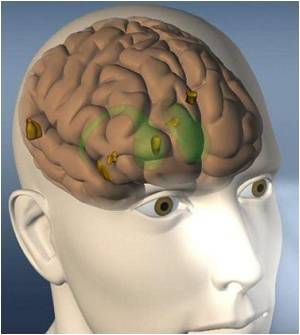
The way in which key circuits in the brain control movement has been shown by scientists at the Gladstone Institute of Neurological Disease (GIND) and Stanford University.

The research team was led by GIND investigator Anatol Kreitzer, PhD, who collaborated with Stanford's Karl Deisseroth, MD PhD, creator of a light activation technology that enabled scientists to activate specific circuits in the motor regions of brain.
"Scientists had identified and diagrammed these circuits in the late 80s and early 1990s, but there had been no way to test their function in animal models," explained Dr. Kreitzer. "This research used genetic methods to allow mice to produce a light-sensitive protein in very select group of cells in the brain."
For decades, a leading theory predicted that our movements are controlled by "go" and "stop" circuits, or pathways, that exert a sort of push-pull control over motor function. Signals are sent to the motor control center in the brain cortex to say, "Yes, go ahead and do this," or "No, stop. Don't do this." In Parkinson's disease, these pathways were thought to go out of balance, causing "stop" signals to dominate. But the function of these pathways had never been experimentally tested.
The researchers used a molecular "switch" from green algae called channelrhodopsin-2 (ChR2), which is turned on by blue light. Scientists genetically engineered ChR2 specifically into cells of either the stop or go pathways in a mouse.
A fiber optic the width of a human hair was then inserted into the brain. When a laser connected to the fiber optics was illuminated, the light in the brain caused the ChR2 to turn on, and this action stimulated only the stop cells or only the go cells. When the light was off, the cells were quiet. As soon as the light was turned on, they became active. When the light was turned off, the activity stopped.
Advertisement
Researchers found that the mouse with the fiber optics implanted in the brain moved normally with the laser turned off and froze when the laser was turned on. With the laser off, and the mouse's movement was restored. "It's not something we can do for just a second," Kreitzer said. "We can do this for as long as the laser is on."
Advertisement
"We found that by activating the 'stop' pathway we could mimic Parkinson's disease. But what we really wanted was a strategy to treat the disease symptoms." For this, Dr. Kreitzer and colleagues turned to the "go" pathway. "We thought that by activating the 'go' pathway, we could re-balance these brain pathways and directly restore movement, even in the absence of dopamine." The strategy worked even better than expected. "We generated mice that lacked dopamine, and these mice showed many of the same symptoms found in humans with Parkinson's disease. But when we activated the 'go' pathway in these mice, they began to move around normally again. We restored all of their motor deficits with this treatment, even though the mice still lacked dopamine."
He added that selective stimulation of the motor planning circuitry might be important for treating Parkinson's and also other disorders involving these circuits, such as Huntington's disease, Tourette's syndrome, obsessive-compulsive disorder, and addiction. Finally, by using these methods to identify the important circuits, new drugs can be developed to home in on these specific circuits now that their function is known.
Source-Eurekalert


