A new device called ultra-low field magnetic resonance imaging (MRI) has captured the first blurry shots of a human brain, revealing its activity as well as structure.
A new device called ultra-low field magnetic resonance imaging (MRI) has captured the first blurry shots of a human brain, revealing its activity as well as structure. MRI scanners detect how hydrogen atoms respond to magnetic fields in order to image the human body. They typically require fields of a few tesla – about 10,000 to 100,000 times stronger than the Earth's magnetic field. The powerful magnets necessary make scanners pricey and also dangerous for people with metal implants.
The new method hits a sample with a 30-millitesla magnetic field, about 100 times weaker than is normally used in MRI. The device then uses a stronger 46-microtesla magnetic field – about the same as the Earth's magnetic field – to capture images of the sample."The cost of MRI can be reduced dramatically," New Scientist quoted Vadim Zotev of Los Alamos National Laboratory in New Mexico, US, as saying. The new system employs several ultra-sensitive sensors called super-conducting quantum interference devices (SQUIDs), which have to be kept at very low temperatures.
"The most expensive part of our system is the liquid helium cryostat, which costs about 20,000 dollars," Zotev added. Ultra-low field MRI scanning was first performed with a single SQUID in 2004 by a group led by John Clarke at University of California, Berkeley, US, but this only allowed objects about the size of an apple to be scanned.
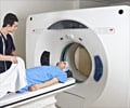
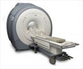
However, the new device uses seven SQUIDs and can scan much larger objects. MRI systems in the clinic today require a patient to be slotted into a long, cylindrical tube. Ultra-low field MRI machines can be much more open.
"Microtesla MRI is more suitable for surgical environment than high-field MRI," Zotev said. "Some medical equipment can be conveniently placed inside [the scanner]," including surgical robots, Zotev added. Today's MRI machines can also be problematic for people with metal implants, since intense magnetic fields can move or heat them causing damage to surrounding tissue.
Experiments have revealed that ultra-low field MRI can image materials even when metal is placed near the magnets (Journal of Magnetic Resonance, vol.179 p.146).
Advertisement
"This is the main advantage of the new set-up. It's a nice step forward," Clarke said.
Advertisement
LIN/P


