Medindia LOGIN REGISTER
Medindia

Migration of New Imaging Technique from Lab to Clinic

by Nancy Needhima on Mar 26 2012 11:10 PM

Each new imaging technology is charismatic since out of the blue it reveals what had been concealed and makes visible what had been invisible.
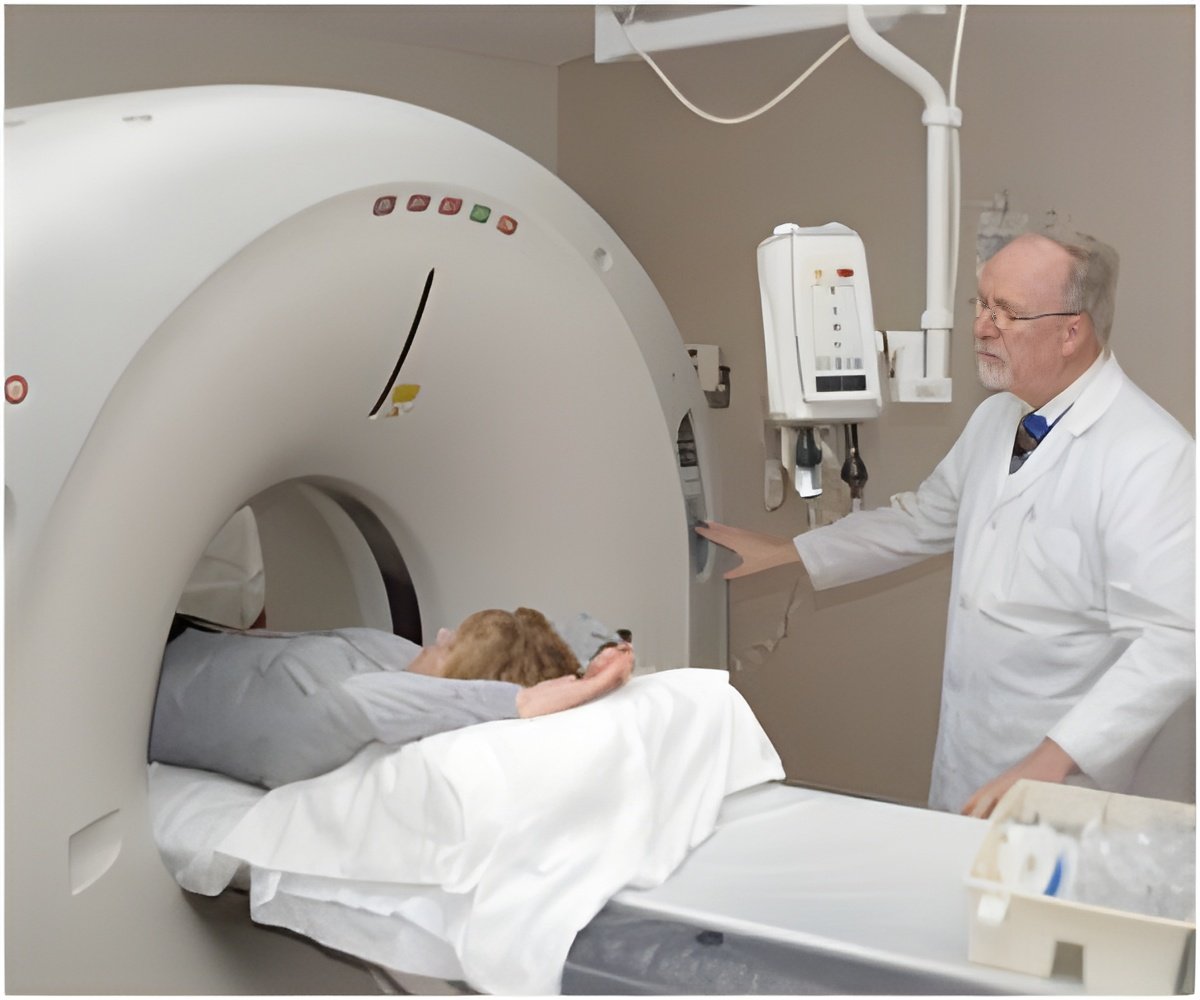
Migration of New Imaging Technique from Lab to Clinic
Each new imaging technology is charismatic since out of the blue it reveals what had been concealed and makes visible what had been invisible. So, too, with photoacoustic tomography, which is allowing scientists to virtually peel away the top several inches of flesh to see what lies beneath.
The technique achieves this depth vision by an elegant marriage between light and sound, combining the high contrast due to light absorption by colored molecules such as hemoglobin or melanin with the spatial resolution of ultrasound.

Lihong V. Wang, PhD, the Gene K. Beare Distinguished Professor of Biomedical Engineering in the School of Engineering & Applied Science at Washington University in St. Louis, summarizes the state of the art in photoacoustic imaging in the March 23 issue of Science.

He is already working with physicians at the Washington University School of Medicine to move four applications of photoacoustic tomography into clinical trials. One is to visualize the sentinel lymph nodes that are important in breast cancer staging; a second to monitor early response to chemotherapy; a third to image melanomas; and the fourth to image the gastrointestinal tract.

Source-Eurekalert


Advertisement

Home

Consult

e-Book

Articles

News

Calculators

Drugs

Directories

Education

Consumer

Professional