Motion analysis as both a diagnostic as well as a monitoring tool since it allows clinicians to objectively measure the gains proper medical or surgical intervention.
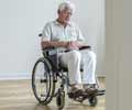
Newswise — At fourteen, cerebral palsy hampered Anthony’s ability to walk. He and his mother feared that within the next few years, he would be confined to a wheelchair.
As is the case with more than 500,000 Americans with this crippling disorder, Anthony was left with few options and his family turned to Hospital for Special Surgery with the hope of finding answers to effectively address his condition.They were introduced to David M. Scher, M.D., a pediatric orthopedic surgeon at Special Surgery, who also serves as co-medical director of the Leon Root, M.D. Motion Analysis Laboratory at the hospital. Dr. Scher believed that motion analysis could be used as an additional imaging tool to figure out the best approach to surgery to prevent Anthony from losing his ability to walk.
“We evaluated Anthony’s functional limitations during walking, a process known as gait analysis,” said Dr. Scher. “We discovered Anthony had severe deformities, specifically in his knee that prevented his ability to walk and the analysis allowed our team to quantify some of the problems with his gait that we saw qualitatively.”
Dr. Scher values motion analysis as both a diagnostic as well as a monitoring tool since it allows clinicians to objectively measure the gains that are achieved following proper medical or surgical intervention.
With the help of gait analysis, Dr. Scher determined which surgical approach was the best option. The surgery entailed cutting the end of the thigh bones to straighten the legs and moving the patellar tendon further down the leg to give it more leverage. Next, Dr. Scher cut the right heel bone to move it in line with his leg and fused a deformed joint in the foot at the bottom of the big toe that prevented Anthony from being able to push off with each step.
Now 16, Anthony’s ability to walk efficiently has been restored. What’s more, Anthony can expect that he will maintain the ability to walk and remain mobile and productive throughout his lifetime.
Advertisement
“Think of motion analysis as having 15 pairs of eyes focused on a moving body part,” said Dr. Scher. “Motion analysis imaging shows all three planes – front, side and top down all at once. While other imaging options can show these views one by one, they can’t show them simultaneously and in motion.”
Advertisement
By focusing on the trunk, pelvis, hips, knees and ankles, digital video cameras take images of a patient’s movement and process the data through a high-tech computer system that analyzes the movement and reveals precisely where muscles and joints are and are not functioning properly.
One design feature of the Leon Root, M.D. Motion Analysis lab is the mechanical isolation of the data collection zone. The laboratory floor is completely detached from the structure of the building it resides in, explained Dr. Hillstrom. Constructing the floor in this fashion was purposefully done to ensure that measurements during motion analysis had a negligible error due to vibrations that occur in most buildings.
The use of high resolution digital video cameras also helps obtain a highly accurate description of each patient’s movement patterns for both clinical and research applications. The air-bearing assemblies beneath each force plate allow them to be adjustably repositioned, further adding to the versatility of the lab. From the gait of a toddler to a seven-foot basketball player, a wide range of neuromusculoskeletal activities may be studied.
Source-Newswise
LIN/M


