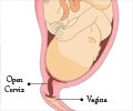
Three-dimensional images of the cervix can help understand the detailed micro-structure of the organ and its associated complications.

‘By developing a detailed image of the cervix, the chances of signs of potential problems before pregnancy can be monitored.’


Around a quarter of miscarriages during the fourth to sixth month of pregnancy (mid-trimester) occur because of weaknesses in the cervix.
The research team at the University of Leeds hope by developing a detailed image of its structure, they can develop ways of monitoring women for signs of potential problems before they become pregnant. Mr. Nigel Simpson, Associate Professor of Obstetrics and Gynaecology, said: "Ultrasound monitoring is used to identify women at risk - where their cervix is unable to support the pregnancy. But little is known about why that problem develops.
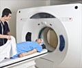
MRI of the Cervix
MRI techniques were used to create 3D images of the cervix. This is the first time extremely high-resolution imaging has been used to understand the detailed microstructure of this organ.
James Nott, from the Faculty of Medicine and Health and lead author, said: "A lot of our understanding of the biology of the cervix is rooted in research carried out 50 years ago. By applying the imaging techniques that have been used in the brain, we can get a much clearer understanding of the tissue architecture that gives the cervix its unique biomechanical properties."
Advertisement
During pregnancy, these fibers provide a strong supporting barrier - keeping the fetus and amniotic sac in place and preventing micro-organisms from entering the uterus.
Mr. Simpson said: "This study's findings have encouraged us to explore new imaging techniques to check the integrity of these fibers before or during pregnancy in order to identify at-risk mums, intervene earlier, and so prevent late pregnancy loss and pre-term birth."
Source-Eurekalert


