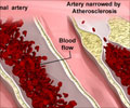
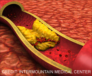
For the first time, researchers can provide early detection of plaques that have a high likelihood of clotting and/or rupture.
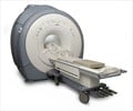
‘Early detection of plaques that have a high likelihood of clotting and/or rupture has been made possible by using non-invasive Magnetic Resonance Imaging (MRI).’


Researchers at BUSM, under the direction of James A. Hamilton, Professor of Physiology and Biophysics and Research Professor of medicine at BUSM, pioneered the use of MRI to identify high-risk plaques in a unique experimental model encompassing both atherosclerosis and thrombosis (atherothrombosis). The group established criteria for distinguishing vulnerable and stable plaques from analysis of in vivo (from a living organism) images of mature plaques. The plaques were then tested to determine their stability.
In the new study, images were obtained at monthly intervals to provide information about the pathways of progression (plaque history) of individual plaques in the vessel and to determine whether MRI can discriminate vulnerable and stable plaques at early times.
Hamilton, who is the corresponding author, said, "Vulnerable plaques and stable plaques showed different physiological progression patterns beginning after one month. Stable plaques exhibited no features of vulnerability at any time, whereas vulnerable plaques showed a progression of vulnerable features, especially in the last month. Successive MRIs could provide a non-invasive means of identifying plaques that are evolving to become a high risk for rupture. This work provides us a unique model for evaluating potential therapies after vulnerable plaques are clearly established."
Source-Eurekalert

