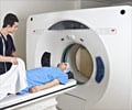
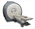
Magnetic Resonance Imaging (MRI) is twice as sensitive as conventional methods to detect heart damage early in patients with the immune system disorder sarcoidosis
According to a study conducted by cardiologists of Duke University Medical Center, Magnetic Resonance Imaging (MRI) can be used as a conventional method for detecting heart damages early in patients with the immune system disorder sarcoidosis, who are at higher risk of dieing from heart problems.
By using MRI to discover minute areas of heart damage before they grow larger, physicians may be able to take action to prevent sudden cardiac death, which is a leading cause of death in patients with sarcoidosis, the researchers said.Sarcoidosis is characterized by the formation of tiny inflammatory growths called granulomas. Although granulomas tend to cluster in the lungs, in lymph nodes and under the skin, they also can form in the heart. When they do, it currently is difficult to determine which patients will develop heart damage, the researchers said.
"We found that MRI was sensitive in detecting small areas of damage in the hearts of patients with sarcoidosis, and we were further able to correlate these areas of damage with future adverse outcomes," said Duke cardiologist Manesh Patel, M.D., who presented the results of the study on Sunday, Nov. 12, at the annual scientific sessions of the American Heart Association, in Chicago. "The MRI technology is very good at obtaining high-resolution images of heart muscle and distinguishing normally functioning heart cells from those that are damaged or destroyed."
The Duke Cardiovascular Magnetic Resonance Center supported the study.
According to Patel, conventional methods identify cardiac damage in only 5 percent to 7 percent of sarcoidosis patients. The standard evaluation includes an electrocardiogram, which is an electrical test of the heart, coupled with one of a number of different cardiac imaging techniques.
But previous studies in which autopsies were performed on sarcoidosis patients indicate that up to 30 percent of such patients exhibit evidence of heart damage, he said.
Advertisement
For their analysis, the Duke researchers identified 81 sarcoidosis patients consecutively referred for evaluation at Duke. All of the patients received a standard clinical evaluation including an electrocardiogram and on average 1.6 non-cardiac MRI imaging tests, and a cardiac MRI scan. The conventional method identified 10 patients (12.3 percent) with heart damage, while the cardiac MRI identified 21 patients (26 percent) with areas of heart damage, Patel said.
Advertisement
The patients in the study were followed by their treating physicians for an average of 13 months, and Patel's team examined their records to see if they had died or had experienced medical problems related to the heart's electrical system.
According to Patel, it is commonly thought that when sarcoidosis damages a portion of heart muscle, the damaged areas can block or reroute the electrical impulses that keep the heart beating. Sudden cardiac death can occur when the heart's internal electrical system is disrupted, causing the heart to beat erratically and, in some cases, to stop.
By the end of the follow-up period, five patients had died from cardiac causes, two had experienced heart-beat abnormalities requiring treatment to bring the heart back into normal rhythm and one needed a pacemaker implanted to maintain normal heart rhythm, Patel said.
The imaging technique that the team used is called delayed enhancement cardiac MRI. In this approach, the researchers inject trace amounts of the element gandolinium into patients before administering the MRI scan.
"Gandolinium is an inert metal, and it cannot enter normally functioning heart muscle cells," Patel said. "However, if small areas of heart muscle are damaged, there are areas that absorb the gandolinium like a sponge. The MRI then detects where the gandolinium accumulates and shows us where the damage is located."
Source-Eurekalert
SRI


