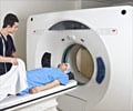
MRI is a noninvasive technique that produces high-quality images of structures and other internal organs inside the body. The technology's applications and image quality have grown steadily since MRI was introduced into medicine in the 1990s.
MRI is a noninvasive technique that produces high-quality images of structures and other internal organs inside the body. The technology's applications and image quality have grown steadily since MRI was introduced into medicine in the 1990s.
It is now possible to tag the work of millions of individual strands of heart muscle fibers, which work together to ensure circulation though out our body, by MRI. The smallest deformations have been mapped inside the hearts of patients who have either silently developed heart disease or remained healthy.Changes in heart mass and volume, linked to early signs of left ventricle problems, can now be identified even before the functionality of the entire heart is lost. Clues have also been obtained regarding the regional development of heart disease and the migration to different parts of the heart and cardiovascular system.
The scan analyzed different parameters of heart function, including thickness of various heart walls, pumping volume, ejection fraction and shortening fraction, blood pressure and body mass index. Deterioration in heart function was observed with changes in the structural pattern of the heart or technically represented as remodeling of the heart.
These results raise the possibility that early treatments for regional heart problems could be instituted to prevent or suppress larger problems from developing. To conclude, it is now possible to map changes of the heart in an early stage of their development using MRI as a key tool in combating cardiovascular disease.


