Laquinimod triggers immune cells within the CNS to produce and release brain-derived neurotrophic factor (BDNF).
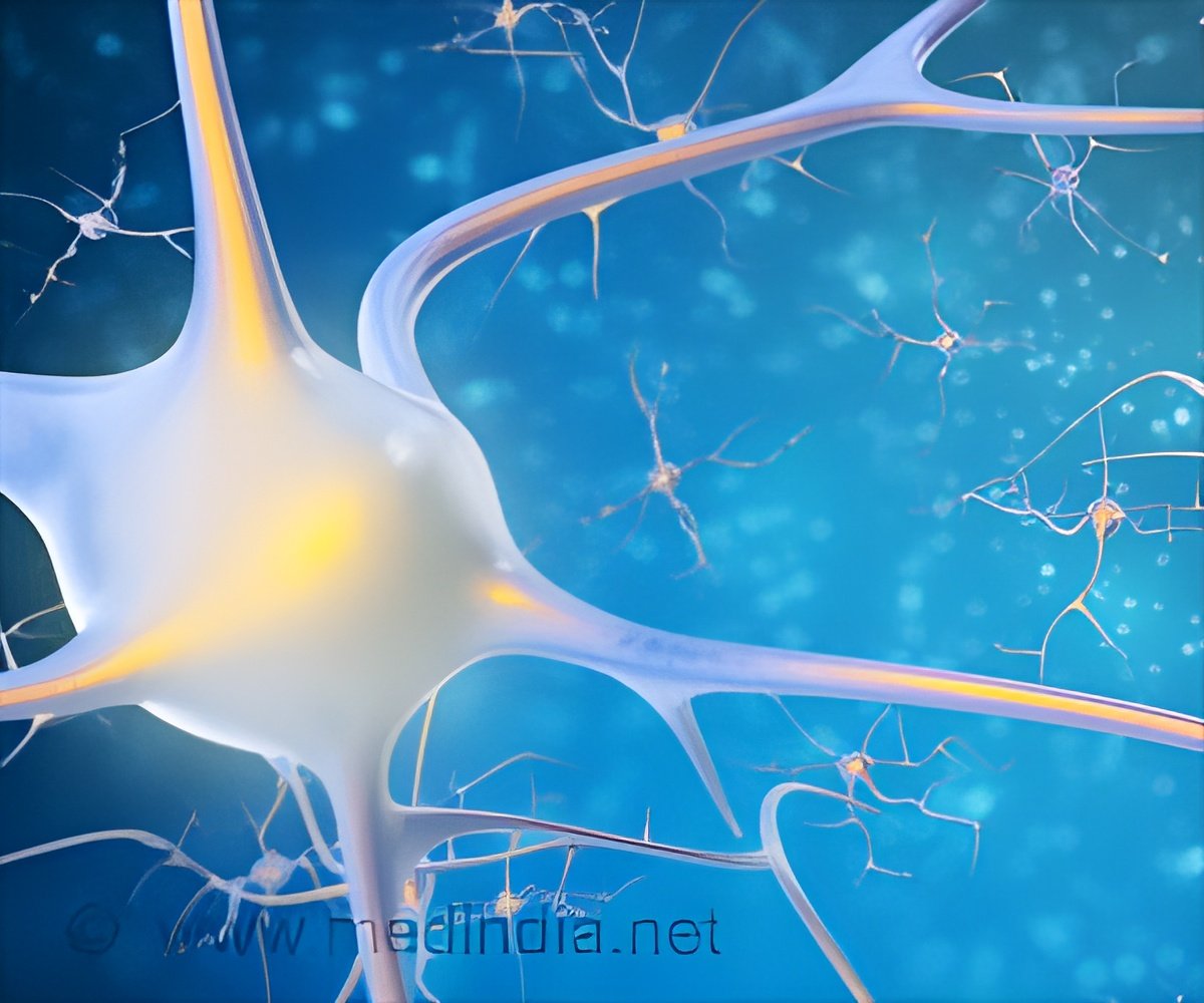
Neurotrophins, such as BDNF, are essential for the development and maintenance of neurons and axons in the central nervous system. Although BDNF is mainly produced by neurons, several types of immune cells also secrete BDNF, suggesting a role in neuroprotection.
To elucidate the mechanism of action of laquinimod, and to explore its potential neuroprotective capacity, the researchers evaluated levels of BDNF in the serum of RRMS patients treated with laquinimod in phase II clinical trials. A significant and robust BDNF increase occurred in 76% of the laquinimod-treated patients, with up to an 11-fold increase in BDNF serum levels observed in individual patients. BDNF elevation in individual patients was independent of relapse rate, and there was no correlation between BDNF levels and age, gender, or baseline disability. Yet, the source of serum BDNF subsequent to treatment remained questionable.
Experiments with animal models corroborated the findings in human patients. Experimental autoimmune encephalomyelitis (EAE; a model of MS) was induced in mice with a conditional BDNF deficiency in immune cells (LLF mice) and in wild-type (WT) control mice. Treatment with laquinimod resulted in a significant reduction in EAE incidence and disease severity in the WT mice. The effect of laquinimod was significantly reduced in the LLF-mice.
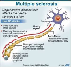
Further studies showed that WT mice treated with a suboptimal dose of laquinimod demonstrated a significant reduction in the inflammatory area and level of demyelination. These mice also displayed a reduction of macrophage infiltration and a significant preservation of axonal densities in comparison with laquinimod-treated LLF mice and controls. The data suggest a BDNF-dependent mechanism of action for laquinimod in autoimmune demyelination.
To investigate whether laquinimod-treated monocytes mediate immune modulation in vivo, laquinimod-stimulated monocytes were injected into WT mice at an early EAE disease stage. The mice showed less severe disease course than controls. Transfer of laquinimod-treated cells derived from LLF mice into WT mice with ongoing EAE did not influence disease course. The cells also secrete significantly less IL-10, an immunomodulatory cytokine that is associated with the generation of regulatory monocytes.
Advertisement
Source-Eurekalert

