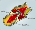
Researchers have developed nanoparticles that aid in the detection of blood clots and also make the clots visible to spectral CT, a new kind of X-ray technology.

"Every year, millions of people come to the emergency room with chest pain. For some of them, we know it's not their heart. But for most, we're not sure," he added.
When there is any doubt, the patient must be admitted to the hospital and undergo tests to rule out or confirm a heart attack.ather than an overnight stay to make sure the patient is stable, this new technology could reveal the location of a blood clot in a matter of hours.
The nanoparticles are used with a new type of CT scanner that is capable of 'seeing' metals in colour.
The new technology, called spectral CT, uses the full spectrum of the X-ray beam to differentiate objects that would be indistinguishable with a regular CT scanner that sees only black and white.
Lanza said the new scanner uses the same principle astronomers use to look at the light from a star and tell what metals it contains.
Advertisement
"Each nanoparticle is carrying a million atoms of bismuth," said Lanza.
Advertisement
Bismuth is a toxic heavy metal, so it can't be injected into the body. Instead, Pan used a compound made of bismuth atoms attached to fatty acid chains that can't come apart in the body.
He then dissolved the compound in a detergent and encapsulated the mixture in a phospholipid membrane.
Much like oil droplets suspended in shaken vinaigrette, these particles self-assemble with the bismuth compound at the core.
Once the nanoparticles carried enough bismuth to be visible to the scanner, Pan added a molecule to the particles' surface that seeks out a protein called fibrin.
Fibrin is common in blood clots but is not found elsewhere in the vasculature.
A spectral CT image with the bismuth nanoparticles targeted to fibrin will provide the same information as a traditional black and white CT image, but the fibrin in any blood clots will show up in a colour, such as yellow or green, solving the problem of calcium interference common to traditional CT scanners.
The spectral CT scanner used in this study is still a prototype instrument. The nanoparticles have only been tested in rabbits and other animal models, but early results show success in distinguishing blood clots from calcium interference.
Source-ANI


