There is a correlation between increased circuit activity in the right side of the brain and the debilitating, involuntary flashbacks triggered by post traumatic stress disorder (PTSD).

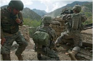
The authorities are being criticized for not doing enough to rehabilitate the returning soldiers. On the other hand, they have to keep up the morale of those being sent to the front.
For those in charge the Minnesota finding could come as a shot in the arm. For the ability to objectively diagnose PTSD through concrete evidence of neural activity, its impact and its manifestation is the first step towards effectively helping those afflicted with this severe anxiety disorder.
Using a technique called Magnetoencephalography (MEG), a non-invasive measurement of magnetic fields in the brain, researchers found differences between signals in the temporal and parieto-occipital right hemispheric areas of the brain among those with PTSD. The temporal cortex, in accordance with earlier findings on the effects of its electrical stimulation during brain surgery, is thought to be responsible for the re-living of past experiences.
The research — led by Apostolos Georgopoulos, M.D., Ph.D., and Brian Engdahl., Ph.D., both members of the Brain Sciences Center at the Minneapolis VA Medical Center and University of Minnesota — is published today in the Journal of Neural Engineering.
It’s a major scientific and medical discovery, Georgopoulos said, because the MEG tests reveal a clear difference in activity among the circuitry in the brains of PTSD sufferers in comparison to those without the condition. The findings are something conventional brain scans such as an X-ray, CT, or MRI have failed to demonstrate.
Advertisement
Besides diagnosing those with PTSD, the researchers also were able to judge the severity of a patient’s suffering, which means the MEG may be able to be used to gauge the how badly patients are impacted by other brain disorders.
Advertisement
Also of particular interest to the scientists: during the study, the brains of PTSD sufferers were in the hyperactive state despite the lack of any immediate external stimulation, evidence found through trial subjects being purposefully put into a “task-free state.” This finding is significant because it confirms that PTSD sufferers can relive terrifying memories at any moment regardless of what they are doing.
“Remarkably, the differences we found between the PTSD and the control groups were documented in a task-free state. Without evoking traumatic experiences, and therefore, reflects the status of steady-state neuronal interactions,” Georgopoulos said.
The trial involved 80 subjects with confirmed PTSD, many of whom suffer the affliction following military service in Vietnam, Afghanistan and Iraq; 18 subjects in PTSD remission; and 284 healthy subjects.
All participants were required to wear the MEG helmet while fixating on a spot 65 cm in front of them for 60 seconds.
The research is one further step in the attempt to ‘biomark’ PTSD, particularly as the results gathered from subjects in remission followed a similar but less pronounced pattern to those with PTSD confirmed as their primary diagnosis, in contrast to the healthy subjects.
This research was funded by the U.S. Department of Veterans Affairs.
Source-Medindia


