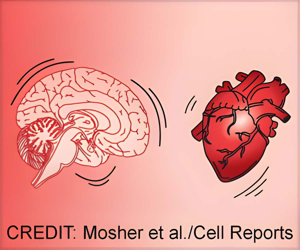
The brain jiggles when the heart beats and now, scientists have identified a new way to use that motion to better study the differences between types of neurons.

‘The tiny brain motion caused by the heartbeat allows scientists to measure the waveform shape more accurately.’


But as the team zoomed in more, they realized that the neurons weren't firing in a different pattern; instead, the brain was jiggling. For every heartbeat, the brain pulses, and the neurons shift their place slightly within the skull. The scientists estimate that the neurons shift about three micrometers, which is less than the width of a hair, during a heartbeat. The appearance of a difference in neuronal firing was created by this movement. "We started from something that many people viewed as a result of brain motion rather than neural activity. They consider it noisy. They consider it as a limitation of their experiment," says Costas Anastassiou, of the Allen Institute, who is the senior author of the study together with Ueli Rutishauser, of Cedars-Sinai Medical Center. "What we were able to show is that if used it in a smart way, this naturally occurring motion of the brain can tell us much more about the identity of the cells we're recording from. This is because measuring the activity of the same neuron from different locations in the brain provides additional information about the neuron."
Conventionally, scientists classify neurons based on their waveform, a characteristic pattern of electrical activity that each neuron emits every time it becomes active, i.e. when it "spikes." The shape of each neuron's waveform is different. By examining the width of the waveform, scientists can reliably categorize neurons into two types: those with narrow and those with broad waveforms.
As the distance between a neuron and the electrode changes, the measured waveform changes as well. By measuring these changes, the team showed that they can differentiate between three different classes of neurons in the human hippocampus: narrow spike (NS), broad spike one (BS1), and broad spike two (BS2). And each class has different firing properties: the researchers found that BS1 neurons coordinate their activity with gamma waves, whereas BS2 neurons coordinate their activity with theta waves.
"Gamma and theta waves are patterns of activity in the brain that are highly relevant to cognition. We know, for example, that memory and learning are very closely linked with theta oscillations. We know that attention is closely linked with gamma oscillations," says Anastassiou.
Advertisement
One of the challenges in neuroscience is that there is often a difference between how neurons behave in living humans and how they behave when investigated in isolation in a brain slice. Through recordings from human brain tissue, the researchers were able to construct single-cell models that simulate the biophysical features and morphology of real neurons. The model bridges the in-vivo brain and ex-vivo brain slice recordings to serve as a novel tool to categorize neurons. The computational models of human neurons can be used to better understand the signals we record from live human beings implanted with electrodes.
Source-Eurekalert


