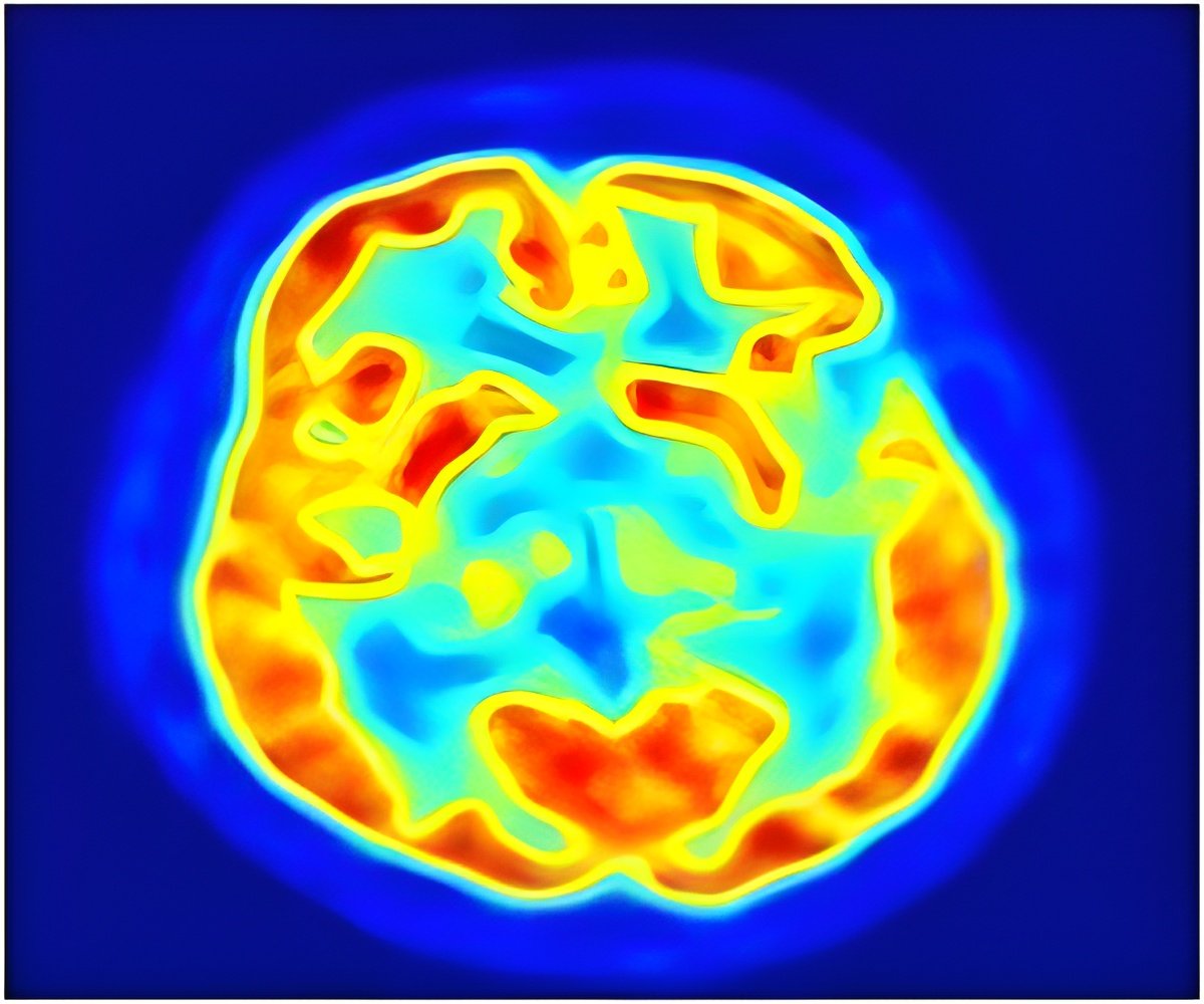
A new approach for measuring early brain development in infants that leads to more accurate whole brain growth charts.

For the first time, researchers used magnetic resonance imaging (MRI) of the newborn brain to calculate the volume of multiple brain regions and to map out regional growth trajectories during the infant's first 90 days of life. The study followed the brain growth of full term and premature babies with no neurological or major health issues.
"A better understanding of when and how neurodevelopmental disorders arise in the postnatal period may help assist in therapeutic development, while being able to quantify related changes in structure size would likely facilitate monitoring response to therapeutic intervention. Early intervention during a period of high neuroplasticity could mitigate the severity of the disorders in later years," said Dominic Holland, PhD, first author of the study and researcher in the Department of Neurosciences at UC San Diego School of Medicine.
For more than two centuries, clinicians have tracked brain growth by measuring the outside of the infant's head with a measuring tape. The results are then plotted on a percentile chart to indicate if normal growth patterns exist. While the measurement is helpful for observing growth, it does not reveal if the individual structures within the brain are developing normally.
On average, researchers found the newborn brain grows one percent each day immediately following birth but slows to 0.4 percent per day by three months. In general for both sexes, the cerebellum, which is involved in motor control, grew at the highest rate, more than doubling volume in 90 days. The hippocampus grew at the slowest rate, increasing in volume by only 47 percent in 90 days, suggesting that the development of episodic memory is not as important at this stage of life.
"We found that being born a week premature, for example, resulted in a brain four to five percent smaller than expected for a full term baby. The brains of premature babies actually grow faster than those of term-born babies, but that's because they're effectively younger – and younger means faster growth," said Holland. "At 90 days post-delivery, however, premature brains were still two percent smaller. The brain's rapid growth rates near birth suggest that inducing early labor, if not clinically warranted, may have a negative effect on the infant's neurodevelopment."
Advertisement
Next steps involve continuing to make advances in the application of different MRI modalities to examine the newborn brain. MRI provides high quality images of different types of tissue and does not involve radiation, like computed tomography (CT). Future research will investigate how brain structure sizes at birth and subsequent growth rates are altered as a result of alcohol and drug consumption during pregnancy.
Advertisement
Source-Eurekalert


