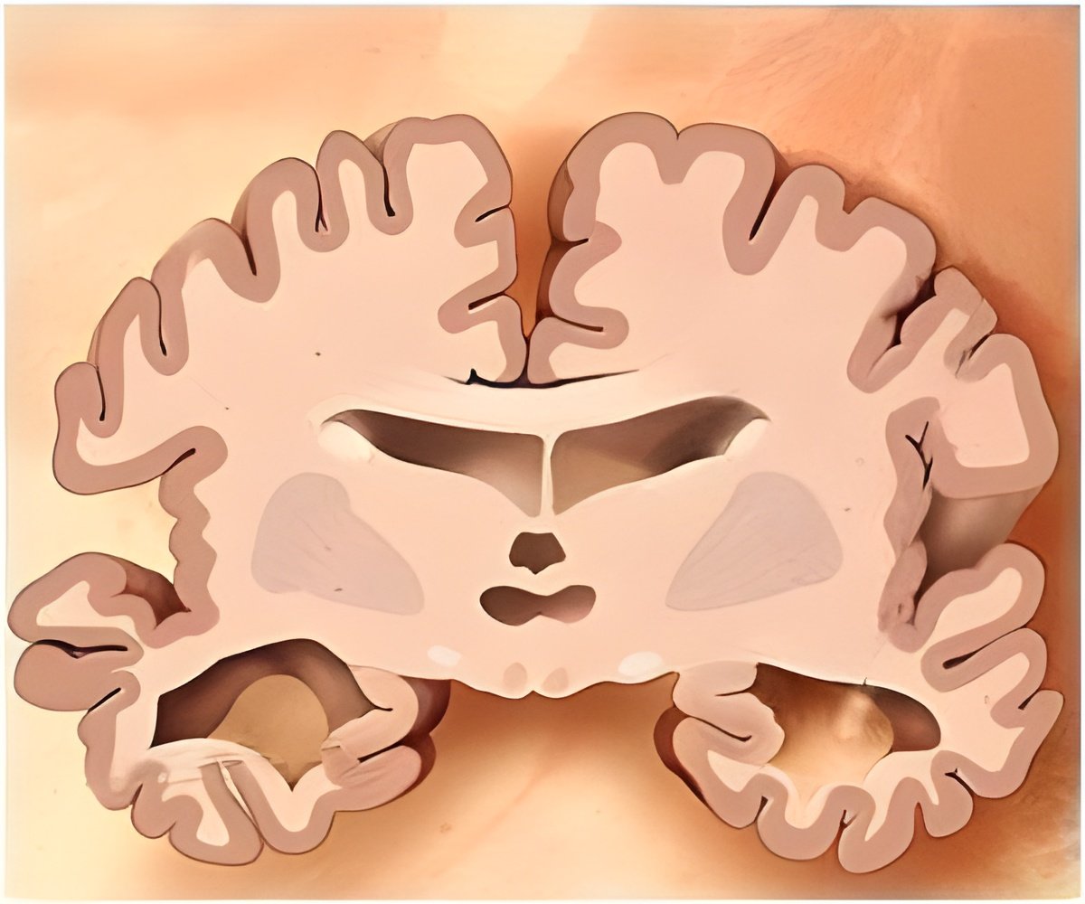
Designing therapies to combat and monitor the disease by early detection of damage to the brain would be possible by the use of non-invasive MRI probe.

The accumulated toxins have associated magnetic nanostructures and they help show up as dark areas in MRI scans of the brain.
"We have a new brain imaging method that can detect the toxin that leads to Alzheimer's disease," said neuroscientist William L Klein who led the research team along with materials scientist Vinayak P Dravid.
"Using MRI, we can see the toxins attached to neurons in the brain. We expect to use this tool to detect this disease early and to help identify drugs that can effectively eliminate the toxin and improve health," Klein said.
The conventional imaging method is by positron emission tomography using probes that target amyloid fibrils but these fibrils are not closely linked to the development of the disease, said the study.
The new technology is detecting something different from conventional technology: toxic amyloid beta oligomers instead of plaques, which occur at a stage of Alzheimer's when it is useless to give therapeutic intervention. It is believed that Amyloid beta oligomers is the the culprit in the onset of Alzheimer's disease and subsequent memory loss.
Advertisement
"Non-invasive imaging by MRI of amyloid beta oligomers is a giant step forward towards diagnosis of this debilitating disease in its earliest form," said Dravid, the Abraham Harris Professor of Materials Science and Engineering at the McCormick School of Engineering and Applied Science.
Advertisement
Source-Medindia


