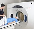
A new technique for conducting cardiac magnetic resonance imaging (MRI) test that improves patient comfort, shortens testing time and has the potential to increase diagnostic accuracy and reliability.
‘A new technique for conducting cardiac magnetic resonance imaging (MRI) test that improves patient comfort, shortens testing time and has the potential to increase diagnostic accuracy and reliability.’


"Our solution is like making a video instead of a still image," Christodoulou said. "MR Multitasking continuously acquires image data and then, when the test is completed, the program separates out the overlapping sources of motion and other changes into multiple time dimensions."
The Nature Biomedical Engineering study shows that MR Multitasking was tested in 10 healthy volunteers and 10 cardiac patients and was found to be accurate, said Debiao Li, PhD, director of the Biomedical Research Institute and the primary investigator of the study. It also was more comfortable for patients, he said, because patients were no longer required to hold their breaths, and the team was able to complete three cardiac MRI tests in as little as 90 seconds--where current standard approaches would have taken many times as long.
"Now we collect all the data throughout the entire test and sort it afterwards," Li said. "We get full control after the test, as opposed to trying to control the body's natural movement during imaging." Incorporating motion and time into the MR Multitasking analysis means that the images have six dimensions, Christodoulou said. "If a picture is 2-D, then a video is 3-D because it adds the passage of time," Christodoulou said. "Our videos are 6-D because we can play them back four different ways: We can playback cardiac motion, respiratory motion and two different tissue processes that reveal cardiac health." MR Multitasking is already in use at several medical centers in the U.S. and China, Li said. The team now is looking to expand MR Multitasking to patients with other diseases, like cancer.
"People have to breathe no matter what disease they have, so the ability to separate out the motion is important to many medical specialties," Li said.
Source-Eurekalert

