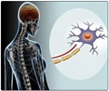
Purdue University researchers say that new imaging technique, called coherent anti-Stokes Raman cattering microscopy (CARS), can be helpful in early detection of multiple sclerosis.
Purdue University researchers say that new imaging technique, called coherent anti-Stokes Raman cattering microscopy (CARS), can be helpful in early detection of multiple sclerosis.
The researchers have studied and recorded how myelin degrades real-time in live mice using the technique.Myelin is the fatty sheath coating the axons, or nerve cells, that insulate and aid in efficient nerve fibre conduction. In diseases such as multiple sclerosis, the myelin sheath has been found to degrade.
The researchers insist that the unprecedented feat of looking real-time at the actual progress of demyelination will advance understanding of multiple sclerosis, and perhaps promote early detection of the disease.
During the course of study, the researchers injected a compound called lysophosphatidylcholine (LPC) into the myelin of a mouse.
When the researchers used CARS to observe the condition of the mice thereafter, they saw an influx of calcium ions into the myelin.
Boffins now believe that this influx may be behind the beginning of the process of myelin degradation.
Advertisement
LIN/C


