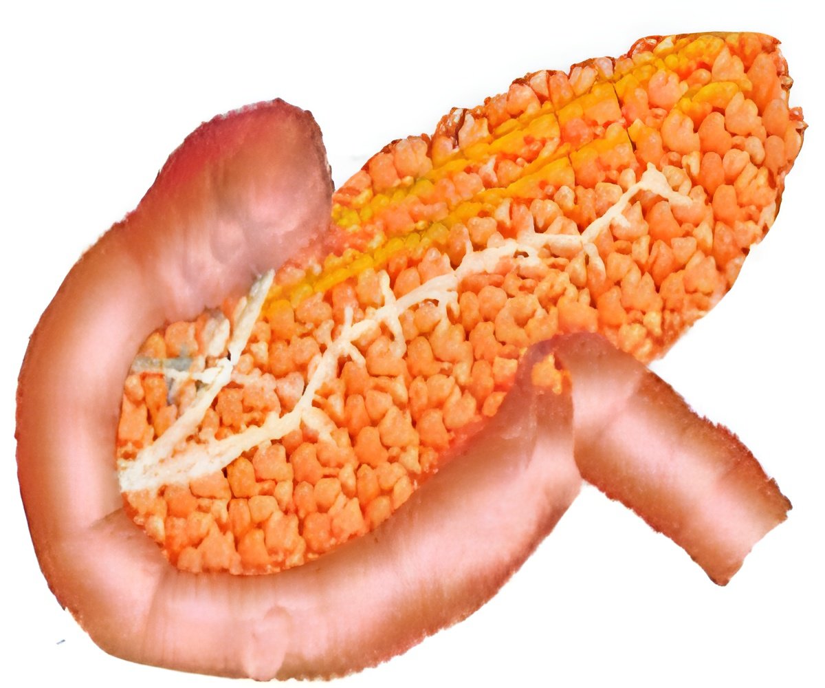
Near-infrared imaging system can reliably distinguish between pancreatic cysts that are low-risk and high-risk for becoming malignant.

To test the diagnostic potential of OCT imaging, researchers from four Boston-area institutions led by Nicusor Iftimia from Physical Sciences, Inc. used the technique to examine surgically removed pancreatic tissue samples from patients with cystic lesions.
By identifying unique features of the high-risk cysts that appeared in the OCT scans, the team developed a set of visual criteria to differentiate between high and low risk cysts.
They then tested the criteria by comparing OCT diagnoses to those obtained by examining thin slices of the pancreatic tissue under a microscope.
Their results showed that OCT allowed clinicians to reliably differentiate between low-risk and high-risk cysts with a success rate close to that achieved by microscope-assisted examinations of slices of the same samples.
The study has been described in the August issue of the Optical Society's (OSA) open-access journal Biomedical Optics Express.
Advertisement


