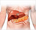
Optical technology developed by a Northwestern University biomedical engineer shown to be effective in the early detection of colon cancer now appears promising for detecting pancreatic cancer
Optical technology developed by a Northwestern University biomedical engineer shown to be effective in the early detection of colon cancer now appears promising for detecting pancreatic cancer, the fourth most common cause of cancer deaths in the United States.
Known as a silent killer, with no method of early detection, pancreatic cancer spreads rapidly and seldom is detected in its early stages. The new technique could lead to the first screening method for pancreatic cancer in asymptomatic patients, said Vadim Backman, developer of the technology and professor of biomedical engineering at Northwestern’s Robert R. McCormick School of Engineering and Applied Science.Backman and Yang Liu, a former graduate student of Backman’s, teamed up with physicians at Evanston Northwestern Healthcare (ENH) to test the technique in a pilot study of 51 patients. The researchers found they could detect both early- and advanced-stage pancreatic cancer without touching or imaging the pancreas.
The extraordinarily sensitive technique, which is minimally invasive and takes advantage of certain light-scattering effects, can detect abnormal changes in cells lining the duodenum even though the cells appear normal when examined with a conventional microscope. The results, which will be published in the Aug. 1 issue of the journal Clinical Cancer Research, show that the changes accurately predict the presence of cancer.
More than 30,000 people in the United States die each year from pancreatic cancer. Count Basie, René Magritte, Billy Carter and Joseph Cardinal Bernardin all died from it; Luciano Pavarotti is fighting the disease. The overall five-year survival rate is less than 5 percent; most patients die within the first two years. If detected early, when the tumor can be successfully removed, however, the survival rate is 100 percent if a precancerous lesion is found and 50 percent for a stage 1 cancer.
“Using endoscopy and taking biopsies of the pancreas are extremely risky procedures that are not used on asymptomatic patients,” said Backman. “When a patient becomes symptomatic, it is too late. This creates a vicious cycle that we want to break.
“We have found that we can take measurements safely in the duodenum and use a biological phenomenon called the ‘field effect’ to our advantage,” he said. “If you have a precancerous or cancerous lesion in the pancreas, even tissue that looks normal and is away from the lesion -- including in the duodenum, a different organ than the pancreas -- will have molecular and other kinds of abnormal changes. No one can detect these changes earlier than we can.”
Advertisement
The researchers found that the same optical markers that were significant in earlier colon cancer studies at ENH using Backman’s technology proved also to be significant for pancreatic cancer. An optical marker is a signature at the sub-micro level that shows changes in tissue due to the presence of a precancerous lesion or cancer.
Advertisement
Most cancers in the pancreas originate from the main pancreatic duct, a 10-centimeter-long duct located in the pancreas that perforates the duodenum, the first and shortest part of the small intestine. The pancreatic duct is difficult to reach and if attempted, it is a risky procedure with a 20 percent chance of significant complications, including acute pancreatitis.
In the study, biopsies of normal-looking tissue were taken from the duodenum near the opening of the pancreatic duct for analysis. For each sample, light is shined on the tissue. The light scatters and some of it bounces back to sensors in the fiber-optic probe. A computer analyzes the pattern of light scattering, looking for the “fingerprint” of carcinogenesis in the nanoarchitecture of the cells.
The researchers found the technique identified with 100 percent accuracy each person who had a resectable cancerous tumor in the pancreas. (Resectable means the tumor can be removed surgically, which in this study is defined as stage 1 or 2 tumors.) Some people were identified who did not have a tumor; it is uncertain whether this is a false finding or if it means those people could be at risk for developing pancreatic cancer and need to be watched closely.
The method combines two complementary technologies developed by Backman and colleagues in his lab: four-dimensional elastic light-scattering fingerprinting (4D-ELF) and low-coherence enhanced backscattering spectroscopy (LEBS). The researchers found that the two combined work better than one alone in pancreatic cancer screening.
The success of the pancreatic cancer screening study follows on the heels of extremely positive results in studies using the two optical technologies for the early detection of colon cancer. In the colon cancer work, Backman has been collaborating with ENH gastroenterologist Hemant Roy, M.D., associate professor of medicine at the Feinberg School, who is overseeing clinical trials at Evanston Hospital. (Roy also is a collaborator on the pancreatic cancer work.)
“The results in our colon cancer work, in which measurements are taken from the rectum, led us to wonder if we could use tissue taken from the duodenum to screen for pancreatic cancer,” said Backman. “Our study published in Clinical Cancer Research has shown that not only can we detect large tumors but early tumors as well.”
“This new work extends the concept of the field effect, or field carcinogenesis, to the pancreas,” said Roy. “While the pancreatic cancer research is preliminary, this extraordinarily exciting work offers the prospect of providing an accurate and practical means for screening this lethal malignancy.”
Source-Eurekalert
LIN/C


