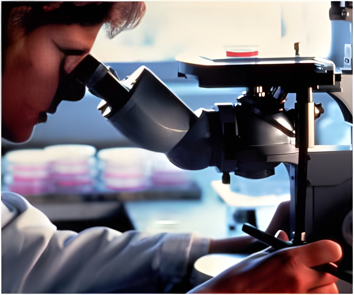
Microscopes, used to see the single living cells are now being used to diagnose illness in hard-to-reach areas of the body.

Dr. Kahaleh often threads a tiny microscope into the narrow bile ducts that connect the liver to the small intestine to hunt for cancer.
He also uses the device to minutely explore the pancreatic duct as one of a few doctors in the country to use such technology in this way.
But because these devices are comparatively new, Dr. Kahaleh, chief of endoscopy at the Center for Advanced Digestive Care at NewYork-Presbyterian/Weill Cornell and professor of clinical medicine at Weill Cornell Medical College, suspected that the specialists who are beginning to use them might be interpreting what they see in different ways.
That's exactly what he and his research team discovered, when they sent six different specialists at five different medical institutions recorded videos taken by a probe-based confocal laser endomicroscopy (pCLE) deep inside 25 patients with abnormally narrowed bile ducts.
The study demonstrated that there was "poor" to "fair" agreement on the clinical significance of what the physicians were viewing in the videos - whether what they saw represented cancer, simple inflammation, or a benign condition.
Advertisement
"We can see detail that was just unimaginable a decade ago - this breakthrough is born for the bile duct and those tiny tubes and complicated organ structures that no one has ever been able to visualize before.
Advertisement
The study was published in Digestive Diseases and Sciences.
Source-ANI


