Researchers were able to prompt a new period of plasticity, or capacity for change, in the neural circuitry of the visual cortex of juvenile mice in a new study.
Researchers were able to prompt a new period of "plasticity," or capacity for change, in the neural circuitry of the visual cortex of juvenile mice in a new study.
The scientists believe that the approach might some day be used to create new periods of plasticity in the human brain that would allow for the repair of neural circuits following injury or disease.The strategy involved transplanting a specific type of immature neuron from embryonic mice into the visual cortex of young mice.
It could be used to treat neural circuits disrupted in abnormal foetal or postnatal development, stroke, traumatic brain injury, psychiatric illness and aging.
Like all regions of the brain, the visual cortex undergoes a highly plastic period during early life.
In mice, this critical period of plasticity occurs around the end of the fourth week of life.
The catalyst for the so-called critical period plasticity in the visual cortex is the development of synaptic signalling by neurons that release the inhibitory neurotransmitter GABA.
Advertisement
In the study, the scientists wanted to see if the embryonic neurons, once they had matured into GABA-producing inhibitory neurons, could induce plasticity in mice after the normal critical period had closed.
Advertisement
Then they transplanted the MGE cells into the animals' visual cortex at two different juvenile stages.
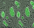
The cells, targeted to the visual cortex, dispersed through the region, matured into GABAergic inhibitory neurons, and made widespread synaptic connections with excitatory neurons.
The scientists then carried out a process known as monocular visual deprivation, in which they blocked the visual signals to one eye in each of the animals for four days.
When this process is carried out during the critical period, cells in the visual cortex quickly become less responsive to the eye deprived of sensory input, and become more responsive to the non-deprived eye, creating alterations in the neural circuitry.
This phenomenon, known as ocular dominance plasticity, greatly diminishes as the brain matures past this critical postnatal developmental period.
The researchers found that the transplanted cells' impact occurred once they had reached the cellular age of inhibitory neurons during the normal critical period.
The finding suggests that the normal critical period of plasticity in the visual cortex is regulated by a developmental program intrinsic to inhibitory neurons, and that embryonic inhibitory neuron precursors can retain and execute this program when transplanted into the postnatal cortex, thereby creating a new period of plasticity.
"The findings suggest it ultimately might be possible to use inhibitory neuron transplantation, or some factor that is produced by inhibitory neurons, to create a new period of plasticity of limited duration for repairing damaged brains. It will be important to determine whether transplantation is equally effective in older animals," said author Dr. Sunil P. Gandhi, PhD.
The study has been published in the journal Science.
Source-ANI
RAS


