The micromechanics occurring within each cell, and the complex relationship between a cell and its cell wall, are yet to be understood fully.

‘Cell biomechanics plays a significant role in plant development. University of Vermont researchers have developed a method that promises to shed light on single cell biomechanics by capturing individual cells in microscopic gel beads.’


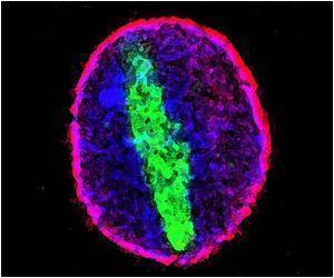
The beads are no wider than a strand of hair, a mere sixty micrometers, but they allow researchers to manipulate the external environment of a single cell and study how the cell responds. They are made using agarose, a material that maintains a fluid state at warm temperatures and hardens as it cools. "We're enthusiastic about this method being a useful tool for researchers interested in mechanical signaling at the cellular level," says Matthew S. Grasso, a graduate student working in Dr. Philip Lintilhac's laboratory in the Plant Biology Department. The new microbead protocol is available in a recent issue of Applications in Plant Sciences.
The first step in creating the microbeads is to prepare the protoplasts from plant tissue. For this study, Grasso used a tobacco cell line. Within a developed piece of plant tissue the cells would look much like a grid, with the grid lines being the cell walls. To get a close look at the mechanics within each cell, Grasso first strips the cells of their cell walls, creating a suspension of free-floating, membrane-enclosed plant protoplasts.
"In the plant body, cells are subject to the mechanical forces generated by their own cell walls, as well as by the cells that surround them. Using individual protoplasts helps control these variables, making it easier to interpret how cells respond to a given mechanical stimulus," says Grasso.
Cells are constantly communicating with each other via signals that pass through cell walls. Recent studies have uncovered a bit about these chemical signals, but the micromechanics occurring within each cell, and the complex relationship between a cell and its cell wall, are yet to be understood fully.
Advertisement
"Unraveling the nuances of the droplet microfluidics system took some time. For a while, it was confusing as to what the different variables were, making it difficult to control them and achieve consistency," says Grasso. Dr. Rachael Oldinski in the Mechanical Engineering Department at the University of Vermont provided assistance and specialized laboratory equipment for the development of the bead protocol.
Advertisement
Within twenty-four hours of bead formation, the membrane-enclosed plant protoplasts regenerate their cell walls. From there, the cells expand and multiply, bursting the beads open. Observing this regenerative ability of the cells under highly controlled conditions could reveal unprecedented knowledge of cellular function.
Source-Eurekalert


