The new approach suggests how the X-ray beams generated by CT can be measured in a way that allows scans from different devices to be usefully compared to one another.
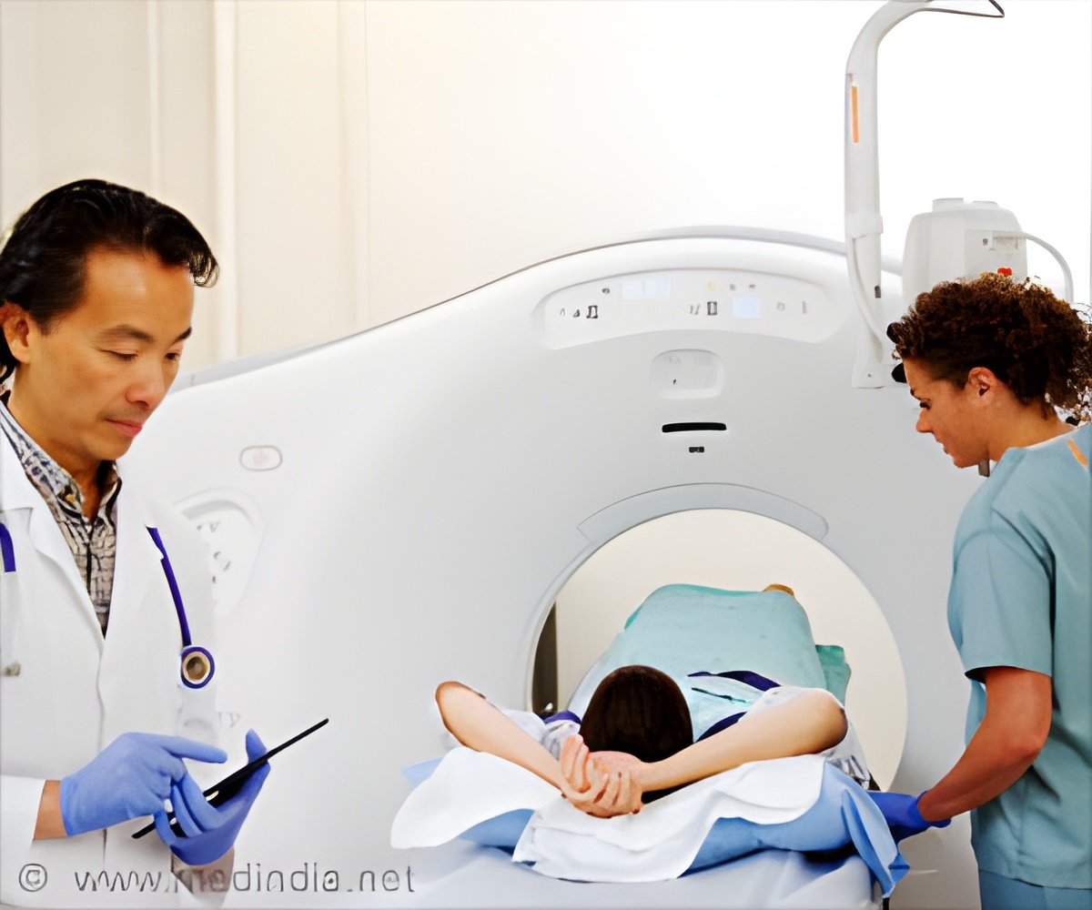
‘New approach offers a pathway to create the first CT measurement standards connected to the International System of Units (SI) by creating a more precise definition of the units used in CT--something the field has lacked.’


"If the technical community could agree on a definition, then?the vendors could create measurements that are interchangeable," said NIST's Zachary Levine, a physicist and one of the paper's authors. "Right now, calibration is not as thorough as it could be." An object's ability to block X-rays--its "radiodensity"--is measured in Hounsfield Units (HUs), named for the Nobel Prize winning co-inventor of CT. Calibration of a CT machine, something every radiology facility has to perform regularly, involves scanning an object of known radiodensity called a phantom and checking whether these measurements give the right number of HUs.
A problem is that a CT scanner's tube--essentially its X-ray generating "light bulb"--creates a beam that is the X-ray version of white light, full of photons with different wavelengths that correspond to their energy. (If the human eye could see X-rays, you could run the tube's beam through a prism and see it break into a spectrum of colors.) Because a photon's penetrating power depends on its energy, the beam's overall effect on the phantom has to be averaged out, making it challenging to define the calibration.
Further complicating the situation is the way the tube's X-ray light has to change depending on the type of scan. Denser body parts need more penetrating X-rays, so the tube has a sort of color switch allowing its operator to adjust the tube voltage to match the job. Adjusting the tube's voltage alters the spectrum of the beam, so that it ranges between something like a "cool white" and a "warm white" light bulb. The variable spectrum makes it tougher to ensure that the calibration is correct for all voltages.
Add these complications to the differences that exist among various CT machine manufacturers, and you get a lot of trouble for anyone who wants to link the calibration of any given scanner to a universal standard. But if it could be done, there would be far-reaching benefits to both industry and medicine.
Advertisement
Better calibration could make diagnosis more efficient and less costly as well, Levine said.
Advertisement
The NIST team had to overcome the uncertainties created by the tube's broad X-ray spectrum and tube voltage setting. Their idea was to fill several phantoms with different concentrations of powdered chemicals that are common in the body, and compare the phantoms' radiodensity using CT. The comparison would help link HUs to the number of moles per cubic meter, which are both SI units.
"Executing this idea was tricky, because the volume of a mole depends on the size of a given chemical molecule," Levine said. "A mole of salt takes up more space than a mole of carbon, for example. And the air in the powders represented a further complication."
The trickiness would make all but a math aficionado wince: Each chemical in the mixture could be characterized by two numbers, but the entire phantom created a 13-dimensional space that complicated the data analysis. Fortunately, the team was able to use a linear algebra technique well-known to data science to simplify the data down to two dimensions, which was far more manageable.
"Basically, we've shown that you can create a CT scanner performance target that any design engineer can hit," Levine said. "Manufacturers have been getting different answers in their machines for decades because no one told their engineers how to handle the X-ray spectrum. Only a small change to existing practice is required to unify their measurements."
Source-Eurekalert

