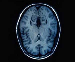
A portable, noninvasive and direct approach is devised to image infant brain activity in a clinical setting without relying on scanning machines.

‘At birth, babies' brains have 100 billion neurons. As a baby grows, so do these neurons, forming branches that connect with other neurons to transmit signals and share information.’


The device consisted of a custom-designed combination video-electroencephalography (EEG) and ultrasound probe weighing only 40 grams (about as heavy as a golf ball), which Charlie Demene and colleagues used to map out subtle changes in blood flow inside small brain vessels correlated with electronic signals of neural activity. Measurements from the probe distinguished between quiet and active sleep (two well-defined mental states) in six healthy napping newborns. The researchers also applied their set up to monitor brain activity in two infants with drug-resistant seizure disorders, which enabled them to detect waves of neurovascular changes spreading throughout the babies' cortexes and trace the brain regions where the seizures originated.
According to the authors, the new technique's low-cost and ease-of-use might make it the standard choice for bedside brain imaging in neonates.
Source-Eurekalert


