The gold standard for detecting advanced fibrosis is a liver biopsy, but the procedure is invasive, and the results are subject to variable interpretation.

‘3D magnetic resonance elastography is probably the most accurate non-invasive method to detect advanced fibrosis in the liver.’


In a paper published in The American Journal of Gastroenterology, researchers at University of California, San Diego School of Medicine conducted a prospective study of 100 patients (56 percent women) with biopsy-proven NAFLD to assess the efficacy of two-dimensional magnetic resonance elastography (MRE) and a novel 3D version. They found that both MRE technologies were highly accurate for diagnosing advanced fibrosis, with 3D perhaps providing additional capabilities in some patients. "3D MRE is probably the most accurate non-invasive method to detect advanced fibrosis," said Rohit Loomba, MD, the study's first author and director of the NAFLD Research Center at UC San Diego School of Medicine.
The researchers say the findings are encouraging because diagnosing NAFLD can be challenging. Current noninvasive techniques, such as molecular biomarkers in blood, are not sufficiently accurate for routine clinical use. Ultrasound-based methods have high failure rates, particularly in obese patients.
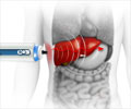
MRE is a specialized version of magnetic resonance imaging (MRI) that propagates mechanical shear waves in liver tissue. An algorithm creates images that quantitatively measure tissue stiffness -- an indicator of fibrosis. The 2D version of MRE is already commercially available and easily implemented on basic MRI systems in clinics. Three-dimensional MRE is more technically demanding and not yet widely available.
Loomba said the prospective study is the first to evaluate 3D-MRE for diagnosing advanced fibrosis in NAFLD patients. Both 2D and 3D were highly accurate, he said. The former was simpler to use, the latter offered improved assessment of spatial patterns and the ability to diagnose larger volumes of tissue. Unlike ultrasound, MRE accuracy did not appear to be impacted by obesity.
Advertisement
Source-Eurekalert


