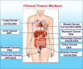
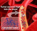
Most cancer deaths result from the spread of the cancer cells from their original locations called as primary tumors.
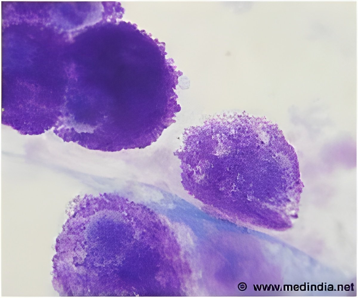
However, not all CTCs in a given patient are alike. Recent discoveries have shown that CTCs are highly heterogeneous - with individual cancer cells possessing very different molecular characteristics - and that only a small subset of these cells actually possess the metastatic potential to spread the disease throughout the body.
Current technologies exist that allow these circulating cells to be captured from the blood of cancer patients, but they are not well equipped to differentiate between the various CTCs present in the blood sample. Instead, they simply count the number of CTCs in a patient sample, rather than identifying the cells that possess the highest metastatic potential. As a result, these tools are less than ideal as they are only able to provide general information on the levels of CTCs rather than a more focused understanding of the disease and its aggressiveness.
Researchers at the Leslie Dan Faculty of Pharmacy at the University of Toronto have developed a new device that provides a way to visualize the heterogeneity of CTCs, and have published their findings in the leading Chemistry journal Angewandte Chemie. Using nanoparticles to tag cells, this device sorts the CTCs collected in a sample into discrete subpopulations based on the phenotype of the cells, and provides a snapshot of the nature of the tumour cells present in patients' blood.
"Recognizing that characterizing the phenotype of circulating tumour cells is more useful for cancer management than quantitating the cells present in a blood sample, we set out to devise a method that would allow us to capture and distinguish between these cells," notes Professor Shana Kelley of the Leslie Dan Faculty of Pharmacy. "In the lab, we were able to demonstrate that the tool was not only highly effective at differentiating these cells, but also proved to be more sensitive than the current leading methods of cellular sorting."
Partnering with collaborators at the Sunnybrook Health Sciences Centre and the London Health Sciences Centre, the researchers collected samples from prostate cancer patients to test the efficacy and ability of the diagnostic platform.
Advertisement
While this study only involved a small number of patients, further validation is planned with several other cancers, including breast, colon, ovarian, lung, and pancreatic cancer.
Advertisement
"As a result, we are excited to pursue new research opportunities in an effort to more accurately and less invasively diagnose and improve the health outcomes for cancer patients."
Source-Eurekalert

