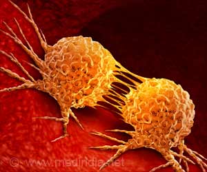
A protein important in embryo formation has been discovered in malignant melanoma cells by researchers of Northwestern University.
A protein important in embryo formation has been discovered in malignant melanoma cells by researchers of Northwestern University.
Scientist discovered this protein called Nodal by injecting malignant melanoma cells in developing zebrafish embryos. The scientists were able to induce abnormal embryonic skull and backbone formation following the injection. The results have been published in online issue of the journal, Nature Medicine."This finding highlights the convergence of tumorigenic and embryonic signaling pathways. From a translational perspective, Nodal signaling provides a novel target for treatment of aggressive cancers such as melanomas," said Mary J. C. Hendrix, the corresponding author, of Children's Memorial Research Center where the discovery was made.
Hendrix is president and scientific director of the Children's Memorial Research Center, professor of pediatrics at Northwestern University Feinberg School of Medicine and a member of the executive committee of The Robert H. Lurie Comprehensive Cancer Center of Northwestern University. Jolanta M. Topczewska and Lynne-Marie Postovit, from Children's Memorial Research Center, co-led the study. Working with Brian Nickoloff of the Cardinal Bernardin Cancer Center at Loyola University Stritch School of Medicine, the investigators found that Nodal protein was present in 60 percent of cutaneous (skin) metastatic melanoma tumors but is absent in normal skin.
They also found that blocking Nodal signaling reduced melanoma cell invasiveness, as well as cancer cell colony formation and tumor-forming ability. Strikingly, nodal inhibition promoted the reversion of these cells toward a normal skin cell type. Like embryonic stem cells, malignant tumor cells similarly receive and send molecular cues during development that promote tumor growth and metastasis, or cancer spread.
The Northwestern study takes advantage of these similarities by using the developing zebrafish to "detect" tumor-derived chemical signals.
In addition, one of the hallmarks of aggressive cancer cells, including malignant melanoma, is their unspecified, "plastic" nature, which is similar to that of embryonic stem cells, expressing genes characteristic of multiple cell types, including endothelial, neural and stem cells.
Advertisement
In this study, the researchers showed that aggressive tumor cells, particularly melanoma, are capable of responding to microenvironmental factors as well as influencing other cells via epigenetic (other than genetic) mechanisms, a quality known as bi-directional cellular communication. Bi-directional cellular communication is integral to both cancer progression and embryological development.
Advertisement
These results also highlight the propensity of tumor cells to communicate bi-directionally and survive within an embryonic microenvironment. Further, the findings illuminate the remarkable plasticity of melanoma cells and the utility of the developing zebrafish as a model for studying the epigenetic modulation of tumor cells.
Melanoma is one of the deadliest forms of cancer. The five-year survival rate for melanoma patients with widespread disease is between 7 percent and 19 percent.
Source: Eurekalert


