Imaging findings of in situ pulmonary artery thrombosis linked to radiation therapy are distinct from acute pulmonary emboli and do not seem to embolize, reports a new study.
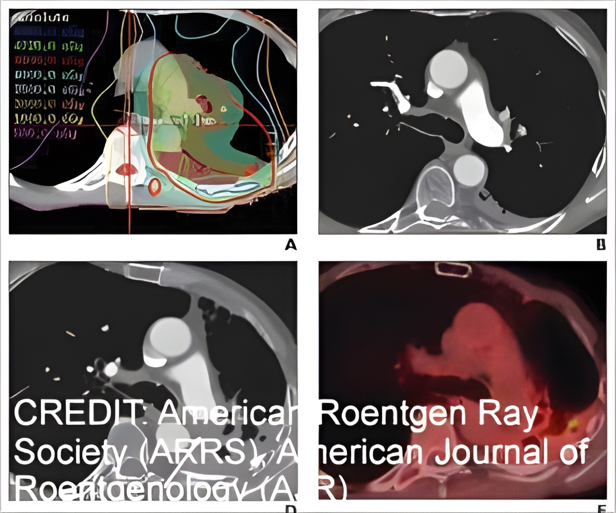
Searching the radiology database of a large teaching hospital to identify patients who had PAT develop after receiving RT, first author Jitesh Ahuja from the thoracic imaging department at the University of Texas' MD Anderson Cancer Center recorded the PAT's CT characteristics: number, location, the appearance of filling defects, as well as the presence of associated lung fibrosis. The terminology (in situ thrombosis vs. acute or chronic pulmonary embolism) is used to describe PAT, the time between completion of RT and development of PAT, the size change of PAT, and any observation of new thrombi emboli on follow-up imaging were also recorded.
With a study population consisting of 27 patients (19 men and eight women) at a mean age of 71 (range, 54-90 years), the primary malignancy was lung cancer in 22 patients (81%) and mesothelioma in five patients (19%). Whereas most PATs were solitary (93%) and nonocclusive (96%) and formed an obtuse angle to the vessel wall (89%), all PATs were eccentric within the involved pulmonary artery and located within the RT volume. The time from completion of RT to PAT's initial diagnosis on CT ranged from 53 to 2,522 days (mean, 675 days). In all patients, CT findings of radiation-induced lung fibrosis were present in the lung supplied by the affected pulmonary artery.
"On follow-up imaging, none of the patients were observed to have filling defects develop in other parts of the PA, which would have suggested embolization," Ahuja et al. added.
Acknowledging that RT is a "key choice" in the multimodality treatment of intrathoracic malignancies, the authors noted that RT-associated cardiovascular complications remain the leading non-cancer-related cause of morbidity and mortality among cancer survivors. "Radiologist awareness of PAT can facilitate accurate diagnosis and impact management," they concluded.
Source-Eurekalert

