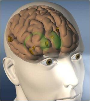
Researchers at Washington University School of Medicine in St. Louis have developed a new technique that provides rapid access to brain landmarks, which have until now been available

The technique makes it possible for scientists to map myelination, or the degree to which branches of brain cells are covered by a white sheath known as myelin in order to speed up long-distance signaling.
"The brain is among the most complex structures known, with approximately 90 billion neurons transmitting information across 150 trillion connections," says David Van Essen, PhD, Edison Professor and head of the Department of Anatomy and Neurobiology at Washington University.
"New perspectives are very helpful for understanding this complexity, and myelin maps will give us important insights into where certain parts of the brain end and others begin," he added.
Easy access to detailed maps of myelination in humans and animals also will aid efforts to understand how the brain evolved and how it works, according to Van Essen.
Neuroscientists have known for more than a century that myelination levels differ throughout the cerebral cortex, the gray outer layer of the brain where most higher mental functions take place. Until now, though, the only way they could map these differences in detail was to remove the brain after death, slice it and stain it for myelin.
Advertisement
The study was recently published in the Journal of Neuroscience.
Advertisement


