Citing recent advances in implantable sensor technology and cartilage scaffolding systems as major developments in the use of engineered cartilage for bone and joint repair are researchers.

Newer, smaller sensing devices that more accurately measure stress loads on joints are giving researchers testing newly grown engineered cartilage within a joint a better understanding of the healing process. Armed with these data, doctors could advise patients on safer, more beneficial levels of activity following joint surgery.
The sensors also transmit their measurements wirelessly, enabling patients undergoing cartilage growth therapy to monitor their own joint stress loads in real time.
The advances appear this month in the Journal of the American Academy of Orthopaedic Surgeons.
The article, "Implantable Sensor Technology: From Research to Clinical Practice," by Eric Ledet, Darryl D''Lima, Peter Westerhoff, John Szivek, Rebecca Wachs and Georg Bergmann, appears in the June 2012 issue of the journal, which publishes research focused on improving the care of patients with musculoskeletal disorders. The article is a review that describes advances in the monitoring of implantable sensors for orthopaedic applications.
Accurately measuring the loads within a repaired joint helps determine smarter ways to get joints to heal, said article co-author John Szivek, professor in the department of orthopaedic surgery at the University of Arizona and director of the UA Orthopedic Research Laboratory in Tucson, Ariz. Szivek also chairs the UA Biomedical Engineering Graduate Interdisciplinary Program.
Szivek said he''s been published with a very elite group of researchers in this particular area of biomedical engineering. "There are only a handful of research groups in the world doing this type of work, and my lab is the only one in the world collecting direct measurements from native tissues," he said.
Advertisement
"It''s a stroke of genius to combine tissue engineering and improved implant measurement technology," said Jennifer Barton, head of the UA biomedical engineering department. "No one really knew what the specific loads were on these joints before this," she said.
Advertisement
"The idea of having a device that''s designed specifically for a patient, tied to a system that provides dynamic feedback directly to that patient, has tremendous possibilities," Barton said.
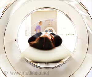
Another advance in the ability of researchers to successfully use cartilage growth for joint repair is the use of computerized tomography, or CT, scans, which are computer-generated medical images used in the diagnosis of tumors and cancer. The 3-D images produced by CT scans are now being used to create patient-specific implantable scaffold systems that support joint cartilage while the tissue grows and gains strength.
Using a 3-D scan of a patient''s joint to custom build an implantable scaffold to support new cartilage growth -- as well as an implanted sensor that provides real-time activity monitoring for the rehabilitated patient -- represent major milestones in cartilage tissue engineering.
"This group has developed a methodology for building an implant that''s closer to the native tissue than anything that has been made before," Barton said.
Source-Newswise


![Genetically Modified Food / Genetically Modified Organism [GMO] Genetically Modified Food / Genetically Modified Organism [GMO]](https://images.medindia.net/patientinfo/120_100/Genetically-Modified-Food.jpg)


