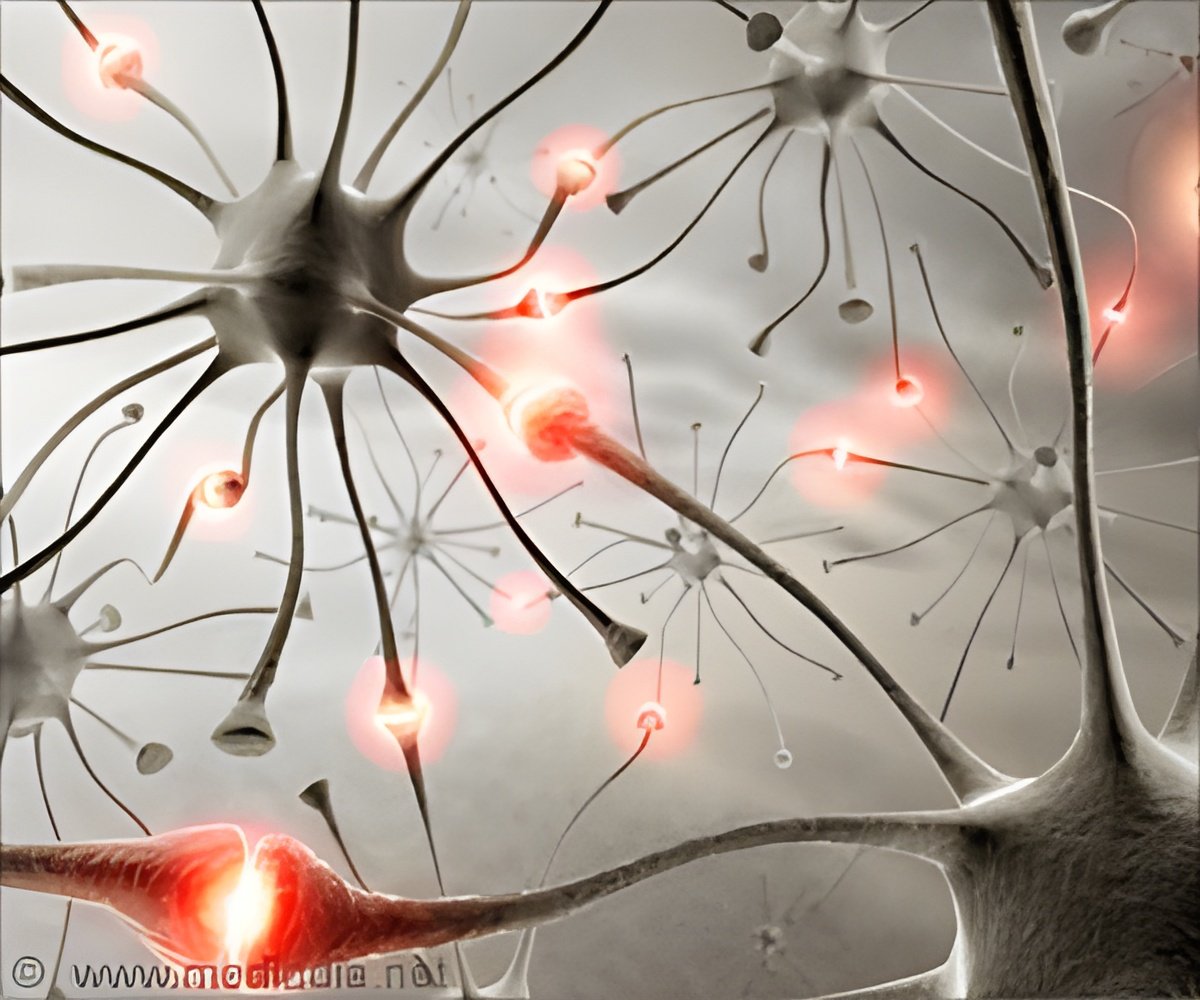
A decades-long debate about how the brain is modified when an animal learns has been solved by researchers at Cold Spring Harbor Laboratory.

‘Changes in neural activity that accompany learning was observed by researchers. They noted that just like our own neurons, dopamine-releasing neurons in the fly are involved in reward and punishment.’


Due to the relative simplicity of fruit fly neural anatomy - there are just two synapses separating odor-detecting antenna from an olfactory-memory brain center called the mushroom body - the diminutive insects have provided a powerful model organism for studying learning. Historically, researchers have monitored neurons in the mushroom body, as well as others to which they send signals, using a technique called calcium imaging. This approach enabled previous researchers to observe changes in neural activity that accompany learning. However, this technique does not reveal precise how the electrical activity of the neurons is modified, since calcium is not the only ion involved in neuronal signaling.
Additionally, it was unclear how the changes that had been seen were related to the behavior of the animal.
Turner and colleagues at CSHL and the Howard Hughes Medical Institute's Janelia Research Campus were able to zoom in to a particularly important part of the fly brain where they were able to connect neural activity to behavior. Toshihide Hige, the lead author of the paper, used his expertise in electrophysiological recordings to directly examine changes in synaptic strength at this site.
The researchers exposed fruit flies to a specific test odor and a very short time later subjected them to an artificial aversive cue. To do so they fired tiny beams of laser light at dopamine-releasing neurons in the mushroom body that were genetically engineered to become active in response to the light. Turner explains, "Just like our own neurons, dopamine-releasing neurons in the fly are involved in reward and punishment. Presenting the smell of cherries, for example, which is normally an attractive odor to flies, while at the same time stimulating a particular dopamine neuron, trains the fly to avoid cherry odor."
Advertisement
Turner said, "Strikingly, the team found a dramatic reduction in the synaptic inputs upon subsequent presentations of the test odor, but not control odors. This drop reflected the decrease in the attractiveness of the odor that resulted from the learning. The average drop in synaptic strength was around 80% - that's huge. We now have a way of investigating synaptic changes with genetic tools to identify molecules involved in learning and really understand the phenomenon at a level that bridges molecular and physiological mechanisms. That mechanistic level of understanding is going to be really important. It's often at the level of molecules that you see really strong connections between Drosophila and other species, including humans."
Advertisement


