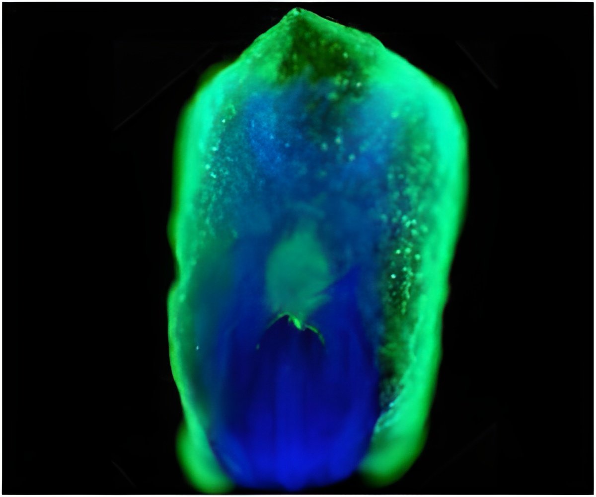
Researchers at the Salk Institute have been able to genetically incorporate unnatural amino acids, such as those emitting green fluorescence, into neural

"Stem cells hold great potential for the treatment of various diseases, yet it has been hard to study how they self renew and produce all of the body's cells," says the study's senior author, Dr. Lei Wang, assistant professor and Frederick B. Rentschler Developmental Chair in the Chemical Biology and Proteomics Laboratory.
"The ability to genetically incorporate unnatural amino acids in stem cell proteins will accelerate our understanding the signaling networks that control these stem cells." he says, Thorough understanding of these mechanisms is critical for safe and reliable stem cell therapeutics.
The study's first author, Dr. Bin Shen, adds that the incorporation of unnatural amino acids "allows researchers to study a particular protein in a living cell or organism, compared to the traditional biochemical methods which are conducted through in vitro settings such as a test tube." These studies can also be conducted in real-time, says Shen, who is a postdoctoral researcher in Wang's lab.
Use of unnatural amino acids (Uaas) was developed by Wang and his colleagues, and was first used in bacteria in 2001, and in mammalian cells in 2007. This is the first report of its use in stem cells.
Salk chemists, molecular and cell biologists, and experts in protein structure collaborated on this study, which was conducted in two stages.
Advertisement
"Current methods for Uaa incorporation are not appropriate for stem cells, because the added genes are often lost before the stem cell has a chance to finish differentiation," Wang says. To solve that problem, the researchers developed a lentiviral-based gene delivery method to incorporate the Uaas into proteins expressed in neural stem cells. "The lentiviral gene therapy technique, which was pioneered by Dr. Inder Verma at Salk, can afford long-lasting expression through stem cell differentiation," Wang says.
Advertisement
"Once you have a stem cell line stably incorporating Uaas, you can custom the Uaa to study stem cell biology," Wang says. "You also get the bonus of obtaining various mature cells with this ability through differentiation, such as neurons, which are difficult to insert Uaas in and expensive to procure in large amounts."
In the first set of experiments, the researchers found out that Uaas were successfully incorporated into neural stem cells, the incorporation lasted through the differentiation, and these cells then produced neurons carrying the fluorescent amino acid.
The second set of experiments was to demonstrate how these Uaas can be used to help solve a biological question. The researchers wanted to know how voltage-sensitive ion channels, which are pore forming proteins, work in neurons. In these nerve cells, membrane ion channels respond to changes in electric current - the charged signal that is passed between neurons - that either activate or silence the neurons.
"We are trying to understand how the electric field of cell membranes can turn on or turn off protein activities - like a switch in a house turns on or off lights," Wang says.
To study this phenomenon, the researchers embedded a fluorescent Uaa into a protein domain that ion channel and other proteins used to sense the electric field in neural stem cells, which produced neurons with the same embedded Uaa. They could then watch, in real time, the fluorescent tag given changes in electrical current across the neuron. "We detected changes in fluorescence intensity of the Uaa when the neurons were stimulated, and these changes are dependent on where the Uaa was incorporated, which hint that different positions of the protein are moving into or outside of the membrane in response to the electric field."
Wang says this experiment, designed to demonstrate the power of Uaas in brain cells, can also be adapted to study other membrane proteins in other cells, no matter where they exist in the body.
Source-Eurekalert


