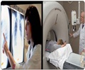
Combined use of two diagnostic imaging techniques -- the so-called CT and PET scans -- can reliably detect early lung cancer, claims a new European
Combined use of two diagnostic imaging techniques -- the so-called CT and PET scans -- can reliably detect early lung cancer, claims a new European study.
Italian researchers from the National Cancer Institute in Milan used both CT (or computed tomography) scans and PET (or positron emission tomography) scans in their study of more than 1,000 heavy smokers. Participants had to have smoked a minimum of 26 cigarettes a day for 37 years.The smokers, all 50 years of age or older, were given low-dose spiral CTs, with or without PET scans, for five years. Lesions up to 5 millimeters were deemed nonsuspicious, and low-dose spiral CT was repeated after 12 months.
By the second year of assessment, 22 participants had been diagnosed with lung cancer. The researchers also detected 440 lung lesions in 298 people, 95 of whom were recalled for high-resolution contrast CT scans.
PET scans were positive in 18 of 20 identified cancer cases, and removal of malignant tissue was successful in 95 percent of the lung cancers, according to results, which are published in the Aug. 23 issue of The Lancet.
"We have shown that low-dose spiral CT combined with selective use of PET can effectively detect early lung cancer," researcher Ugo Pastorino says in a news release. "A more conservative approach to very small CT-detected nodules is justified, and lesions up to 5 millimeters can be followed up at 12 months without any major risks of progression."


