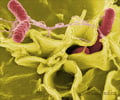
Microbiologists from Oregon State University have developed a novel method to test food for bacterial contamination - something that can revolutionize the food industry.
A novel method to test food for bacterial contamination has been developed by microbiologists from Oregon State University.
They suggest that the new approach would be easier to use, faster and more directly related to toxicity assessment than conventional approaches now used to test food for bacterial contamination and safety.This technique can detect such important food-associated bacteria as Salmonella and Clostridium perfringens, responsible for diarrheal illnesses; Bacillus cereus, responsible for gastrointestinal illness characterized by vomiting and diarrhea, and often referred to as stomach flu, and Clostridium botulinum, which causes toxin-induced botulism, characterized by paralysis.
"Rapid methods are not readily available to directly assess the toxicity of bacterial contamination in a user-friendly fashion," said Janine Trempy, professor of microbiology and associate dean of the OSU College of Science.
"When this new technology is commercially available, we should be able to provide a higher level of assurance to the consumer while avoiding the waste of millions of dollars worth of food that is suspected of bacterial contamination, but actually is safe."
"Bacteria are common on exposed surfaces, including the food products we consume," Trempy said.
"Simply knowing they are there doesn't completely tell you, in a direct measurement, about their potential to make you sick or whether the food is safe to eat," she added.
Advertisement
The researchers found that when these fish encounter certain stressful or threatening environmental conditions, such as exposure to toxic chemicals like mercury, the erythrophores change appearance, and the pigment moves in a characteristic pattern to an internal part of the cell.
Advertisement
Another kind of stressful or threatening situation which also causes the location of pigment to change is the toxic threat posed by illness-causing bacteria. Some of these bacteria are associated with food.
"We discovered that the pigment bearing cells, erythrophores, respond immediately to certain food associated, toxin producing bacteria responsible for making humans sick," Trempy said.
"There is potential to directly assess the toxic behaviour of the contaminating bacteria, not just the simple presence of the DNA or protein of these bacteria.
"And this response can be easily seen under a low-power microscope and quickly quantified, numerically, to describe the intensity of the situation."
Trempy said that it is possible that portable kits could be developed that would not require specialized training to use.
Source-ANI
RAS/SK


