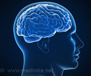
The brain dynamics of a specific region called default mode network, or DMN, are found to be altered by activation of a small brain nucleus in the brainstem.
‘The dynamics of a specific brain region (in the prefrontal cortex) called default mode network, or DMN, are found to be altered by activation of a small brain nucleus in the brainstem — the locus coeruleus (LC).’


The scientific community has long hypothesized the changes to DMN dynamics to be linked with certain behaviors, like those associated with attention deficit-hyperactivity disorder, Alzheimer’s disease, Parkinson’s disease, depression, and autism.However, the precise mechanisms that control DMN dynamics were not understood fully. The present study thereby explored the interplay between neurons and brain chemicals across brain regions, that leads to alterations in DMN dynamics.
Findings of Brain Dynamics
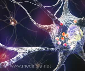
The team used a “chemogenetic” technique, fMRI, and a genetic mouse model (that expresses synthetic receptors in the small brain nucleus in the brainstem — the locus coeruleus [LC]) to study the effects of a neurotransmitter (brain’s chemical messengers) on brain network functional connectivity — a dynamic process crucial for human health and behavior.“Many brain imagers have a vast interest in identifying the circuit mechanisms that control large-scale brain networks. But how a specific neurotransmitter system alters brain-wide dynamics remains incompletely understood. Our work helps explain how norepinephrine (NE) (a neurotransmitter released by LC) affects brain activity and connectivity, leading to changes in the DMN,” says Shih, senior author, and director of the Center for Animal MRI (CAMRI) at the UNC Biomedical Research Imaging Center (BRIC).
It was revealed that chemogenetic activation of LC-NE neurons strengthened the communication of neurons within the frontal cortical regions of the DMN and this functional connectivity was modulated by brain regions — the retrosplenial cortex and hippocampus.
“We believe these two regions potentially could serve as novel targets to control frontal cortical regions and restore DMN function when LC neurons are degenerated in Alzheimer’s and Parkinson’s disease,” says Shih.
Advertisement

