Clues about which protein signaling molecules alert cells to critical changes in their environments, have been unearthed by Johns Hopkins scientists.
The researchers believe that the specific collection of proteins in each cilium, customized to the needs of each cell type, is determined by whether and how cilia keep proteins inside once they enter-not which ones they allow in initially.
"According to our experiments, 90 percent of the proteins in mammalian cells should be able to fit inside cilia based on their size," said Takanari Inoue, Ph.D., assistant professor of cell biology at the Johns Hopkins University School of Medicine.
"But most of them have never been found [inside the cilia], so we think that most proteins do wander in at some point, but only certain ones remain inside," Inoue added.
So-called "primary cilia" have been attracting intense attention as recent research has confirmed their role in monitoring the cell's exterior environment and conveying information to the rest of the cell using an arsenal of signals stored inside the thin interior of each antenna-like cilium.
The results of this study have helped explain how that arsenal is developed.
Advertisement
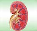
In the kidney, they monitor the flow of urine; in the eye, they sense the wavelength of light; in cartilage, they sense pressure; and in the heart, blood flow. No matter where they are, Inoue said, their job is to translate such mechanical forces-or, in some cases, chemical ones-into molecular signals for the cell, so that it can respond appropriately to its environment.
Advertisement
"A cilium forms like a short drinking straw being pushed outwards from inside an inflated balloon. What we don't know is whether there is some sort of cap over the hole, regulating what goes in and out. We found that more signaling molecules could enter the straw than we thought," explained Inoue.
Previous research in other laboratories suggests that a fixed pore exists at the base of a cilium that only allows relatively small molecules inside. By developing more sensitive experimental methods, the Inoue group was able to show that molecules almost 10 times larger than those known before could enter.
Specifically, they first engineered an anchor-like molecule that selectively embedded itself in the membranes of cilia. On the inside end of the anchor was half of a "molecular snapper." Inside the watery interior of the cell, the team placed fluorescent molecules of known size, fitted with the other half of the molecular snapper. If these fluorescent molecules entered cilia, they would carry their fluorescence with them and be trapped inside when the snappers clicked together, allowing the researchers to easily take images of them.
By repeating this experiment many times with molecules of increasing size, the Inoue team was able to show that every molecular size they tested was able to enter the cilia. The only difference between the molecules of different sizes was their rate of entry: Smaller molecules entered more quickly than larger ones.
Figuring out how cilia select their captives is a question for another study, Inoue noted.
The results of this study will be published online in the journal Nature Chemical Biology.
Source-ANI


